Specification
Catalog# RT1316
Iba1 Recombinant Mouse Monoclonal Antibody [8G8]
-
WB
-
IHC-P
-
FC
-
IP
-
IHC-Fr
-
IF-Cell
-
IF-Tissue
-
Human
-
Mouse
-
Rat
-
Cynomolgus monkey
-
Pig

Overview
Product Name
Iba1 Recombinant Mouse Monoclonal Antibody [8G8]
Antibody Type
Mouse Monoclonal Antibody
Immunogen
Recombinant protein.
Species Reactivity
Human, Mouse, Rat (Predicted: Cynomolgus monkey, Pig)

Validated Applications
WB, IHC-P, FC, IP, IHC-Fr, IF-Cell, IF-Tissue
Molecular Weight
Predicted band size: 17 kDa
Positive Control
Mouse brain tissue, mouse hippocampus tissue, rat brain tissue, RAW264.7, C6, human PBL whole cell lysates.
Conjugation
unconjugated
Clone Number
8G8
Product Features
Form
Liquid
Concentration
1ug/ul
Storage Instructions
Shipped at 4℃. Store at +4℃ short term (1-2 weeks). It is recommended to aliquot into single-use upon delivery. Store at -20℃ long term.
Storage Buffer
PBS (pH7.4), 0.1% BSA, 40% Glycerol. Preservative: 0.05% Sodium Azide.
Isotype
IgG
Purification Method
Protein A affinity purified.
Application Dilution
-
WB
-
1:1,000
-
IHC-P
-
1:500-1:2000
-
IHC-Fr
-
1:1,000
-
IF-Cell
-
1:200
-
FC
-
1:200-1:1,000
-
IP
-
Use at an assay dependent concentration
-
IF-Tissue
-
1:1,000
Applications in Publications
Species in Publications
Target
Function
Ionized calcium-binding adapter molecule 1 (Iba1), also known as allograft inflammatory factor-1 (AIF-1), is a 147 amino acid cytoplasmic, calcium-binding protein that is thought to play a role in macrophage activation and function. Iba1, containing two EF domains, is induced by cytokines and interferons. In an unstimulated state, Iba1 colocalizes with actin, and upon stimulation, translocates to lamellipodia. It is also a marker of human microglia and is expressed by macrophages in injured skeletal muscle. The gene encoding Iba1 maps to chromosome 6p21.33 and resides in the tumor necrosis factor (TNF) cluster of genes located in the region represented by the human major histocompatibility complex (MHC).
Background References
1. Arroba AI et al. Autophagy resolves early retinal inflammation in Igf1-deficient mice. Dis Model Mech 9:965-74 (2016).
2. James RE et al. Loss of galectin-3 decreases the number of immune cells in the subventricular zone and restores proliferation in a viral model of multiple sclerosis. Glia N/A:N/A (2015).
Subcellular Location
Cytoplasm, cytoskeleton.
UNIPROT
Synonyms
AIF 1 antibody
AIF-1 antibody
Aif1 antibody
AIF1 protein antibody
AIF1_HUMAN antibody
Allograft inflammatory factor 1 antibody
Allograft inflammatory factor 1 splice variant G antibody
allograft inflammatory factor-1 splice variant Hara-1 antibody
balloon angioplasty responsive transcription antibody
BART 1 antibody
ExpandAIF 1 antibody
AIF-1 antibody
Aif1 antibody
AIF1 protein antibody
AIF1_HUMAN antibody
Allograft inflammatory factor 1 antibody
Allograft inflammatory factor 1 splice variant G antibody
allograft inflammatory factor-1 splice variant Hara-1 antibody
balloon angioplasty responsive transcription antibody
BART 1 antibody
G1 antibody
G1 putative splice variant of allograft inflamatory factor 1 antibody
IBA 1 antibody
IBA1 antibody
interferon gamma responsive transcript antibody
Interferon responsive transcript 1 antibody
interferon responsive transcript factor 1 antibody
Ionized calcium binding adapter molecule 1 antibody
Ionized calcium-binding adapter molecule 1 antibody
ionized calcium-binding adapter molecule antibody
IRT 1 antibody
IRT1 antibody
Microglia response factor antibody
MRF1 antibody
Protein g1 antibody
CollapseImages
-
Application: IHC-Fr
Species: Mouse
Site: Cerebral cortex
Sample: Frozen section
Antibody concentration: 1/1,000
Antigen retrieval: Not required -
-
-
Application: IHC-Fr
Species: Mouse
Site: Cerebral cortex
Sample: Frozen section
Antibody concentration: 1/1,000
Antigen retrieval: Not required -
Immunohistochemical analysis of paraffin-embedded mouse brain tissue with Mouse anti-Iba1 antibody (RT1316) at 1/500 dilution.
The section was pre-treated using heat mediated antigen retrieval with Tris-EDTA buffer (pH 9.0) for 20 minutes. The tissues were blocked in 1% BSA for 20 minutes at room temperature, washed with ddH2O and PBS, and then probed with the primary antibody (RT1316) at 1/500 dilution for 1 hour at room temperature. The detection was performed using an HRP conjugated compact polymer system. DAB was used as the chromogen. Tissues were counterstained with hematoxylin and mounted with DPX. -
Immunohistochemical analysis of paraffin-embedded mouse hippocampus tissue with Mouse anti-Iba1 antibody (RT1316) at 1/500 dilution.
The section was pre-treated using heat mediated antigen retrieval with Tris-EDTA buffer (pH 9.0) for 20 minutes. The tissues were blocked in 1% BSA for 20 minutes at room temperature, washed with ddH2O and PBS, and then probed with the primary antibody (RT1316) at 1/500 dilution for 1 hour at room temperature. The detection was performed using an HRP conjugated compact polymer system. DAB was used as the chromogen. Tissues were counterstained with hematoxylin and mounted with DPX. -
Immunohistochemical analysis of paraffin-embedded rat brain tissue with Mouse anti-Iba1 antibody (RT1316) at 1/500 dilution.
The section was pre-treated using heat mediated antigen retrieval with Tris-EDTA buffer (pH 9.0) for 20 minutes. The tissues were blocked in 1% BSA for 20 minutes at room temperature, washed with ddH2O and PBS, and then probed with the primary antibody (RT1316) at 1/500 dilution for 1 hour at room temperature. The detection was performed using an HRP conjugated compact polymer system. DAB was used as the chromogen. Tissues were counterstained with hematoxylin and mounted with DPX. -
Immunohistochemical analysis of paraffin-embedded human brain tissue with Mouse anti-Iba1 antibody (RT1316) at 1/500 dilution.
The section was pre-treated using heat mediated antigen retrieval with Tris-EDTA buffer (pH 9.0) for 20 minutes. The tissues were blocked in 1% BSA for 20 minutes at room temperature, washed with ddH2O and PBS, and then probed with the primary antibody (RT1316) at 1/500 dilution for 1 hour at room temperature. The detection was performed using an HRP conjugated compact polymer system. DAB was used as the chromogen. Tissues were counterstained with hematoxylin and mounted with DPX. -
Immunocytochemistry analysis of RAW264.7 cells labeling Iba1 with Mouse anti-Iba1 antibody (RT1316) at 1/200 dilution.
Cells were fixed in 4% paraformaldehyde for 15 minutes at room temperature, permeabilized with 0.1% Triton X-100 in PBS for 15 minutes at room temperature, then blocked with 1% BSA in 10% negative goat serum for 1 hour at room temperature. Cells were then incubated with Mouse anti-Iba1 antibody (RT1316) at 1/200 dilution in 1% BSA in PBST overnight at 4 ℃. Goat Anti-Mouse IgG H&L (iFluor™ 488, HA1125) was used as the secondary antibody at 1/1,000 dilution. PBS instead of the primary antibody was used as the secondary antibody only control. Nuclear DNA was labelled in blue with DAPI.
Beta tubulin (ET1602-4, red) was stained at 1/100 dilution overnight at +4℃. Goat Anti-Rabbit IgG H&L (iFluor™ 594, HA1122) was used as the secondary antibody at 1/1,000 dilution. -
Immunocytochemistry analysis of C6 cells labeling Iba1 with Mouse anti-Iba1 antibody (RT1316) at 1/200 dilution.
Cells were fixed in 4% paraformaldehyde for 15 minutes at room temperature, permeabilized with 0.1% Triton X-100 in PBS for 15 minutes at room temperature, then blocked with 1% BSA in 10% negative goat serum for 1 hour at room temperature. Cells were then incubated with Mouse anti-Iba1 antibody (RT1316) at 1/200 dilution in 1% BSA in PBST overnight at 4 ℃. Goat Anti-Mouse IgG H&L (iFluor™ 488, HA1125) was used as the secondary antibody at 1/1,000 dilution. PBS instead of the primary antibody was used as the secondary antibody only control. Nuclear DNA was labelled in blue with DAPI.
Beta tubulin (ET1602-4, red) was stained at 1/100 dilution overnight at +4℃. Goat Anti-Rabbit IgG H&L (iFluor™ 594, HA1122) was used as the secondary antibody at 1/1,000 dilution. -
Application: IF-Tissue
Species: Mouse
Site: brain
Sample: Paraffin-embedded section
Antibody concentration: 1/1,000 -
Western blot analysis of Iba1 on human PBL whole cell lysate using anti-Iba1 antibody at 1/1,000 dilution.
Please note: All products are "FOR RESEARCH USE ONLY AND ARE NOT INTENDED FOR DIAGNOSTIC OR THERAPEUTIC USE"
Citation
-
Icariin alleviates cognitive dysfunction by reducing neuroinflammation via the cGAS-STING pathway
Author: Fengjuan Lu, Lu Li, Bilian Zheng, Ce Wang, Ziying Liu, Xingshuo Huang, Linyue Song, Chong Ding, Yang Li
PMID: 40403897
Journal: Journal Of Ethnopharmacology
Application: IF
Reactivity: Mouse
Publish date: 2025 May
-
Citation
-
Reduction of D2 receptors on microglia leads to ZBP1-mediated PANoptosis of mPFC in Parkinson's disease depression mice
Author: Qiyue Zhao, Xinyao Zhang, Jing Zhang, Ying Zhang, Lu Jia, Sijia Guo, Mengqing Zhang, Haoran Wang, Yuling Wang, Yue Guan, Yi Zhang, Suibing Miao, Jin-Xia Zhu, Huijie Ma
PMID: 40367691
Journal: International Immunopharmacology
Application: WB,IF
Reactivity: Mouse
Publish date: 2025 May
-
Citation
-
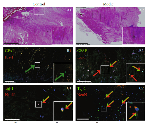
The Effect of Cutibacterium acnes Infection on Nerve Penetration in the Annulus Fibrosus of Lumbar Intervertebral Discs via Suppressing Oxidative Stress
Author: Shan, Z., Wang, X., Zong, W., Li, J., Zheng, B., Huang, B., Zhang, X., Chen, J., & Huang, Y.
PMID: 35265268
Journal: Oxidative Medicine And Cellular Longevity
Application: IF
Reactivity: Rat
Publish date: 2022 Feb
-
Citation
-
Protective effect of scorpion venom heat-resistant synthetic peptide against PM2.5-induced microglial polarization via TLR4-mediated autophagy activating PI3K/AKT/NF-κB signaling pathway
Author: Xin Xu, Henggui Xu, Fei Ren, Lanyi Huang, Jingbin Xu, Fasheng Li
PMID: 33887539
Journal: Journal Of Neuroimmunology
Application:
Reactivity:
Publish date: 2021 Apr
-
Citation
-
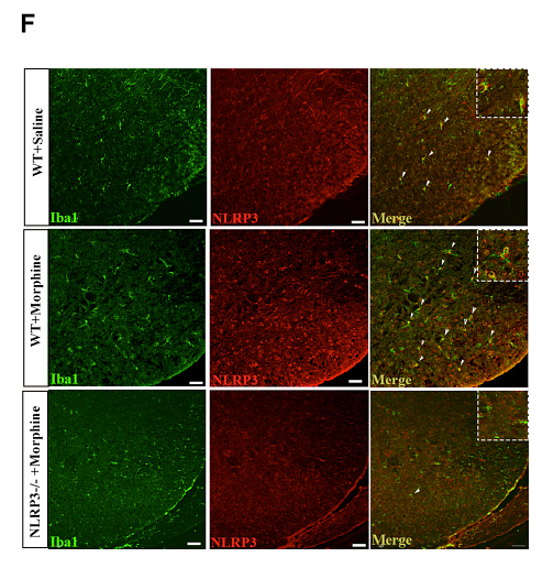
Spinal TLR4/P2X7 Receptor-Dependent NLRP3 Inflammasome Activation Contributes to the Development of Tolerance to Morphine-Induced Antinociception
Author: Tao Xu;Aizhong Wang
PMID: 33061523
Journal: Journal Of Inflammation Research
Application: IF
Reactivity: Mouse
Publish date: 2020 Sep
-
Citation
Alternative Products
Products with the same target and pathway
Iba1 Recombinant Rabbit Monoclonal Antibody [JM36-62] - BSA and Azide free
Application: WB,IHC-P,IHC-Fr,IF-Tissue,IF-Cell,FC,IP
Reactivity: Human,Mouse,Rat,Cynomolgus monkey,Pig
Conjugate: unconjugated
Iba1 Recombinant Mouse Monoclonal Antibody [8G8] - BSA and Azide free
Application: WB,IHC-P,FC,IP,IHC-Fr,IF-Cell,IF-Tissue
Reactivity: Human,Mouse,Rat,Cynomolgus monkey,Pig
Conjugate: unconjugated
Iba1 Recombinant Rabbit Monoclonal Antibody [JM36-62]
Application: WB,IHC-P,IHC-Fr,IF-Tissue,IF-Cell,FC,IP,mIHC
Reactivity: Human,Mouse,Rat,Cynomolgus monkey,Pig
Conjugate: unconjugated
Iba1 Rabbit Polyclonal Antibody
Application: IHC-P
Reactivity: Human,Mouse
Conjugate: unconjugated
Iba1 Recombinant Antibody [JM36-62] - Rat IgG1 (Chimeric) - BSA and Azide free
Application: IHC-Fr,IHC-P
Reactivity: Mouse,Rat,Cynomolgus monkey,Pig
Conjugate: unconjugated
Iba1 Recombinant Antibody [JM36-62] - Guinea pig IgG2 (Chimeric) - BSA and Azide free
Application: IHC-Fr,WB
Reactivity: Human,Mouse,Rat,Cynomolgus monkey,Pig
Conjugate: unconjugated
Iba1 Recombinant Antibody [JM36-62] - Chicken IgY (Chimeric) - BSA and Azide free
Application: IHC-Fr,WB,FC
Reactivity: Human,Mouse,Rat,Pig,Cynomolgus monkey
Conjugate: unconjugated
iFluor™ 488 Conjugated Iba1 Recombinant Rabbit Monoclonal Antibody [JM36-62]
Application: IF-Cell,FC
Reactivity: Human,Mouse,Rat
Conjugate: iFluor™ 488
Iba1 Recombinant Antibody [JM36-62] - Rat IgG1 (Chimeric)
Application: IHC-Fr,IHC-P
Reactivity: Mouse,Rat,Cynomolgus monkey,Pig
Conjugate: unconjugated
Iba1 Recombinant Antibody [JM36-62] - Guinea pig IgG2 (Chimeric)
Application: IHC-Fr,WB
Reactivity: Human,Mouse,Rat,Cynomolgus monkey,Pig
Conjugate: unconjugated
Iba1 Recombinant Antibody [JM36-62] - Chicken IgY (Chimeric)
Application: IHC-Fr,WB,FC
Reactivity: Human,Mouse,Rat,Pig,Cynomolgus monkey
Conjugate: unconjugated