Iba1 Recombinant Rabbit Monoclonal Antibody [JM36-62]


Catalog# ET1705-78
Iba1 Recombinant Rabbit Monoclonal Antibody [JM36-62]
-
WB
-
IHC-P
-
IHC-Fr
-
IF-Tissue
-
IF-Cell
-
FC
-
IP
-
mIHC
-
Human
-
Mouse
-
Rat
-
Pig
-
HA750440
不含抗保成分
-
Cynomolgus monkey
-
unconjugated
Overview
Product Name
Iba1 Recombinant Rabbit Monoclonal Antibody [JM36-62]
Antibody Type
Recombinant Rabbit monoclonal Antibody
Immunogen
Synthetic peptide within N-terminal human Iba1.
Species Reactivity
Human, Mouse, Rat, Pig (Predicted: Cynomolgus monkey)
Validated Applications
WB, IHC-P, IHC-Fr, IF-Tissue, IF-Cell, FC, IP, mIHC
Molecular Weight
Predicted band size: 17 kDa
Positive Control
THP-1 cell lysate, mouse spleen tissue lysate, rat spleen tissue lysate, THP-1, J774A.1, RAW264.7, C6, mouse hippocampus tissue, human kidney tissue, human spleen tissue, mouse brain tissue, rat brain tissue.
Conjugation
unconjugated
Clone Number
JM36-62
RRID
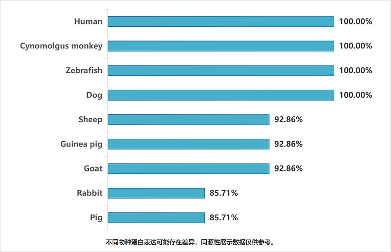
同源性数据
Product Features
Form
Liquid
Concentration
Storage Instructions
Shipped at 4℃. Store at +4℃ short term (1-2 weeks). Store at -20℃ long term.
Storage Buffer
1*TBS (pH7.4), 0.05% BSA, 40% Glycerol. Preservative: 0.05% Sodium Azide.
Isotype
IgG
Purification Method
Protein A affinity purified.
Application Dilution
-
WB
-
1:5,000
-
IHC-P
-
1:1,000
-
IHC-Fr
-
1:1,000-1:2,000
-
IF-Tissue
-
1:500
-
IF-Cell
-
1:250-1:500
-
FC
-
1:1,000
-
IP
-
Use at an assay dependent concentration.
-
mIHC
-
1:2,000
Target
Function
Ionized calcium-binding adapter molecule 1 (Iba1), also known as allograft inflammatory factor-1 (AIF-1), is a 147 amino acid cytoplasmic, calcium-binding protein that is thought to play a role in macrophage activation and function. Iba1, containing two EF domains, is induced by cytokines and interferons. In an unstimulated state, Iba1 colocalizes with actin, and upon stimulation, translocates to lamellipodia. It is also a marker of human microglia and is expressed by macrophages in injured skeletal muscle. The gene encoding Iba1 maps to chromosome 6p21.33 and resides in the tumor necrosis factor (TNF) cluster of genes located in the region represented by the human major histocompatibility complex (MHC).
Background References
1. Hennessy E et al. Systemic TNF-a produces acute cognitive dysfunction and exaggerated sickness behavior when superimposed upon progressive neurodegeneration. Brain Behav Immun 59:233-244 (2017).
2. Arentsen T et al. The bacterial peptidoglycan-sensing molecule Pglyrp2 modulates brain development and behavior. Mol Psychiatry 22:257-266 (2017).
Tissue Specificity
Detected in T-lymphocytes and peripheral blood mononuclear cells.
Post-translational Modification
Phosphorylated on serine residues.
Subcellular Location
Cytoplasm, cytoskeleton, Cell projection, ruffle membrane, Cell projection, phagocytic cup.
Synonyms
AIF 1 antibody
AIF-1 antibody
Aif1 antibody
AIF1 protein antibody
AIF1_HUMAN antibody
Allograft inflammatory factor 1 antibody
Allograft inflammatory factor 1 splice variant G antibody
allograft inflammatory factor-1 splice variant Hara-1 antibody
balloon angioplasty responsive transcription antibody
BART 1 antibody
ExpandImages
-
Application: IHC-Fr
Species: Mouse
Site: Cerebral cortex
Sample: Frozen section
Antibody concentration: 1/2,000
Antigen retrieval: Not required -
Application: IHC-Fr
Species: Rat
Site: Cerebral cortex
Sample: Frozen section
Antibody concentration: 1/1,000
Antigen retrieval: Not required -
Application: IHC-Fr
Species: Mouse
Site: Hippocampus (APP-PS1 mouse)
Sample: Frozen section
Antibody concentration: 1/500
Date by conrtesy of: Mr. Chenxin Ma, School of Basic Medical Sicences, Zhejiang University -
-
-
Fluorescence multiplex immunohistochemical analysis of mouse brain (Formalin/PFA-fixed paraffin-embedded sections). Panel A: the merged image of anti-Iba1 (ET1705-78, Green), anti-Olig2 (ET1604-29, White) and anti-TBR1 (ET1702-97, Red) on brain. HRP Conjugated UltraPolymer Goat Polyclonal Antibody HA1119/HA1120 was used as a secondary antibody. The immunostaining was performed with the Sequential Immuno-staining Kit (IRISKit™MH010101, www.luminiris.cn). The section was incubated in three rounds of staining: in the order of ET1705-78 (1/2,000 dilution), ET1604-29 (1/1,000 dilution) and ET1702-97 (1/1,000 dilution) for 20 mins at room temperature. Each round was followed by a separate fluorescent tyramide signal amplification system. Heat mediated antigen retrieval with Tris-EDTA buffer (pH 9.0) for 30 mins at 95℃. DAPI (blue) was used as a nuclear counter stain. Image acquisition was performed with Zeiss Observer 7 Inverted Fluorescence Microscope.
-
Fluorescence multiplex immunohistochemical analysis of mouse brain (Formalin/PFA-fixed paraffin-embedded sections). Panel A: the merged image of anti-NeuN (ET1602-12, red), anti-Iba1 (ET1705-78, green), anti-GFAP (ET1601-23, gray), anti-Olig2 (ET1604-29, cyan), anti-MAP2 (HA500177, magenta) and anti-CD34 (ET1606-11, yellow) on mouse brain. HRP Conjugated UltraPolymer Goat Polyclonal Antibody HA1119/HA1120 was used as a secondary antibody. The immunostaining was performed with the Sequential Immuno-staining Kit (IRISKit™MH010101, www.luminiris.cn). The section was incubated in six rounds of staining: in the order of ET1602-12(1/5,000 dilution), ET1705-78 (1/2,000 dilution), ET1601-23 (1/5,000 dilution), ET1604-29 (1/1,000 dilution), HA500177 (1/5,000 dilution) and ET1606-11 (1/2,000 dilution) for 20 mins at room temperature. Each round was followed by a separate fluorescent tyramide signal amplification system. Heat mediated antigen retrieval with Tris-EDTA buffer (pH 9.0) for 30 mins at 95℃. DAPI (blue) was used as a nuclear counter stain. Image acquisition was performed with Olympus VS200 Slide Scanner.
-
Application: IF-tissue
Species: Human
Site: Spleen
Sample: Paraffin-embedded section
Antibody concentration: 1/500 -
Application: IF-tissue
Species: Mouse
Site: Cerebral cortex
Sample: Paraffin-embedded section
Antibody concentration: 1/500 -
Application: IF-tissue
Species: Rat
Site: Cerebral cortex
Sample: Paraffin-embedded section
Antibody concentration: 1/500 -
☑ Relative expression (RE)
Western blot analysis of Iba1 on different lysates with Rabbit anti-Iba1 antibody (ET1705-78) at 1/5,000 dilution.
Lane 1: THP-1 cell lysate
Lane 2: HEK-293 cell lysate (negative)
Lane 3: Mouse spleen tissue lysate
Lane 4: Rat spleen tissue lysate
Lysates/proteins at 20 µg/Lane.
Predicted band size: 17 kDa
Observed band size: 17 kDa
Exposure time: 3 minutes;
4-20% SDS-PAGE gel.
Proteins were transferred to a PVDF membrane and blocked with 5% NFDM/TBST for 1 hour at room temperature. The primary antibody (ET1705-78) at 1/5,000 dilution was used in 5% NFDM/TBST at 4℃ overnight. Goat Anti-Rabbit IgG - HRP Secondary Antibody (HA1001) at 1:50,000 dilution was used for 1 hour at room temperature. -
Immunohistochemical analysis of paraffin-embedded mouse brain tissue with Rabbit anti-Iba1 antibody (ET1705-78) at 1/1,000 dilution.
The section was pre-treated using heat mediated antigen retrieval with Tris-EDTA buffer (pH 9.0) for 20 minutes. The tissues were blocked in 1% BSA for 20 minutes at room temperature, washed with ddH2O and PBS, and then probed with the primary antibody (ET1705-78) at 1/1,000 dilution for 1 hour at room temperature. The detection was performed using an HRP conjugated compact polymer system. DAB was used as the chromogen. Tissues were counterstained with hematoxylin and mounted with DPX. -
Immunohistochemical analysis of paraffin-embedded rat brain tissue with Rabbit anti-Iba1 antibody (ET1705-78) at 1/1,000 dilution.
The section was pre-treated using heat mediated antigen retrieval with Tris-EDTA buffer (pH 9.0) for 20 minutes. The tissues were blocked in 1% BSA for 20 minutes at room temperature, washed with ddH2O and PBS, and then probed with the primary antibody (ET1705-78) at 1/1,000 dilution for 1 hour at room temperature. The detection was performed using an HRP conjugated compact polymer system. DAB was used as the chromogen. Tissues were counterstained with hematoxylin and mounted with DPX. -
Immunohistochemical analysis of paraffin-embedded mouse cerebral cortex tissue with Rabbit anti-Iba1 antibody (ET1705-78) at 1/1,000 dilution.
The section was pre-treated using heat mediated antigen retrieval with Tris-EDTA buffer (pH 9.0) for 20 minutes. The tissues were blocked in 1% BSA for 20 minutes at room temperature, washed with ddH2O and PBS, and then probed with the primary antibody (ET1705-78) at 1/1,000 dilution for 1 hour at room temperature. The detection was performed using an HRP conjugated compact polymer system. DAB was used as the chromogen. Tissues were counterstained with hematoxylin and mounted with DPX. -
Immunohistochemical analysis of paraffin-embedded rat cerebral cortex tissue with Rabbit anti-Iba1 antibody (ET1705-78) at 1/1,000 dilution.
The section was pre-treated using heat mediated antigen retrieval with Tris-EDTA buffer (pH 9.0) for 20 minutes. The tissues were blocked in 1% BSA for 20 minutes at room temperature, washed with ddH2O and PBS, and then probed with the primary antibody (ET1705-78) at 1/1,000 dilution for 1 hour at room temperature. The detection was performed using an HRP conjugated compact polymer system. DAB was used as the chromogen. Tissues were counterstained with hematoxylin and mounted with DPX. -
Immunohistochemical analysis of paraffin-embedded human spleen tissue with Rabbit anti-Iba1 antibody (ET1705-78) at 1/1,000 dilution.
The section was pre-treated using heat mediated antigen retrieval with Tris-EDTA buffer (pH 9.0) for 20 minutes. The tissues were blocked in 1% BSA for 20 minutes at room temperature, washed with ddH2O and PBS, and then probed with the primary antibody (ET1705-78) at 1/1,000 dilution for 1 hour at room temperature. The detection was performed using an HRP conjugated compact polymer system. DAB was used as the chromogen. Tissues were counterstained with hematoxylin and mounted with DPX. -
Immunohistochemical analysis of paraffin-embedded human kidney tissue with Rabbit anti-Iba1 antibody (ET1705-78) at 1/1,000 dilution.
The section was pre-treated using heat mediated antigen retrieval with Tris-EDTA buffer (pH 9.0) for 20 minutes. The tissues were blocked in 1% BSA for 20 minutes at room temperature, washed with ddH2O and PBS, and then probed with the primary antibody (ET1705-78) at 1/1,000 dilution for 1 hour at room temperature. The detection was performed using an HRP conjugated compact polymer system. DAB was used as the chromogen. Tissues were counterstained with hematoxylin and mounted with DPX. -
Immunocytochemistry analysis of THP-1 cells labeling Iba1 with Rabbit anti-Iba1 antibody (ET1705-78) at 1/250 dilution.
Cells were fixed in 4% paraformaldehyde for 20 minutes at room temperature, permeabilized with 0.1% Triton X-100 in PBS for 5 minutes at room temperature, then blocked with 1% BSA in 10% negative goat serum for 1 hour at room temperature. Cells were then incubated with Rabbit anti-Iba1 antibody (ET1705-78) at 1/250 dilution in 1% BSA in PBST overnight at 4 ℃. Goat Anti-Rabbit IgG H&L (iFluor™ 488, HA1121) was used as the secondary antibody at 1/1,000 dilution. PBS instead of the primary antibody was used as the secondary antibody only control. Nuclear DNA was labelled in blue with DAPI. Beta tubulin (M1305-2, red) was stained at 1/100 dilution overnight at +4℃. Goat Anti-Mouse IgG H&L (iFluor™ 594, HA1126) was used as the secondary antibody at 1/1,000 dilution. -
Immunocytochemistry analysis of BV2 cells labeling Iba1 with Rabbit anti-Iba1 antibody (ET1705-78) at 1/500 dilution.
Cells were fixed in 4% paraformaldehyde for 20 minutes at room temperature, permeabilized with 0.1% Triton X-100 in PBS for 5 minutes at room temperature, then blocked with 1% BSA in 10% negative goat serum for 1 hour at room temperature. Cells were then incubated with Rabbit anti-Iba1 antibody (ET1705-78) at 1/500 dilution in 1% BSA in PBST overnight at 4 ℃. Goat Anti-Rabbit IgG H&L (iFluor™ 488, HA1121) was used as the secondary antibody at 1/1,000 dilution. PBS instead of the primary antibody was used as the secondary antibody only control. Nuclear DNA was labelled in blue with DAPI. Beta tubulin (M1305-2, red) was stained at 1/100 dilution overnight at +4℃. Goat Anti-Mouse IgG H&L (iFluor™ 594, HA1126) was used as the secondary antibody at 1/1,000 dilution. -
Immunocytochemistry analysis of RAW264.7 cells labeling Iba1 with Rabbit anti-Iba1 antibody (ET1705-78) at 1/250 dilution.
Cells were fixed in 4% paraformaldehyde for 20 minutes at room temperature, permeabilized with 0.1% Triton X-100 in PBS for 5 minutes at room temperature, then blocked with 1% BSA in 10% negative goat serum for 1 hour at room temperature. Cells were then incubated with Rabbit anti-Iba1 antibody (ET1705-78) at 1/250 dilution in 1% BSA in PBST overnight at 4 ℃. Goat Anti-Rabbit IgG H&L (iFluor™ 488, HA1121) was used as the secondary antibody at 1/1,000 dilution. PBS instead of the primary antibody was used as the secondary antibody only control. Nuclear DNA was labelled in blue with DAPI. Beta tubulin (M1305-2, red) was stained at 1/100 dilution overnight at +4℃. Goat Anti-Mouse IgG H&L (iFluor™ 594, HA1126) was used as the secondary antibody at 1/1,000 dilution. -
Immunocytochemistry analysis of C6 cells labeling Iba1 with Rabbit anti-Iba1 antibody (ET1705-78) at 1/250 dilution.
Cells were fixed in 4% paraformaldehyde for 20 minutes at room temperature, permeabilized with 0.1% Triton X-100 in PBS for 5 minutes at room temperature, then blocked with 1% BSA in 10% negative goat serum for 1 hour at room temperature. Cells were then incubated with Rabbit anti-Iba1 antibody (ET1705-78) at 1/250 dilution in 1% BSA in PBST overnight at 4 ℃. Goat Anti-Rabbit IgG H&L (iFluor™ 488, HA1121) was used as the secondary antibody at 1/1,000 dilution. PBS instead of the primary antibody was used as the secondary antibody only control. Nuclear DNA was labelled in blue with DAPI. Beta tubulin (M1305-2, red) was stained at 1/100 dilution overnight at +4℃. Goat Anti-Mouse IgG H&L (iFluor™ 594, HA1126) was used as the secondary antibody at 1/1,000 dilution. -
Flow cytometric analysis of THP-1 cells labeling Iba1.
Cells were fixed and permeabilized. Then stained with the primary antibody (ET1705-78, 1μg/mL) (red) compared with Rabbit IgG Isotype Control (green). After incubation of the primary antibody at +4℃ for an hour, the cells were stained with a iFluor™ 488 conjugate-Goat anti-Rabbit IgG Secondary antibody (HA1121) at 1/1,000 dilution for 30 minutes at +4℃. Unlabelled sample was used as a control (cells without incubation with primary antibody; black). -
Application: Western blot
Species: Pig
Sample: Brain
Antibody concentration: 1/5,000
Date by conrtesy of: Prof. Sen Yan, Guangdong-HongKong-Macau Institutue of CNS Regeneration (GHMICR), Jinan University
Please note: All products are "FOR RESEARCH USE ONLY AND ARE NOT INTENDED FOR DIAGNOSTIC OR THERAPEUTIC USE"
Citation
-
Senolytic therapy ameliorates high-fat diet-induced hippocampal senescence and cognitive decline in mice
Journal: Experimental Neurology
DOI: 10.1016/j.expneurol.2026.115745
IF: 4.2
Application: IF-tissue
Reactivity: Mouse
Publish date: 2026 Mar
-
Giving Old Drugs New Life: Disulfiram Alleviates Microglial Pyroptosis and Disulfidptosis-like Cytoskeletal/Mitochondrial Alterations in Cerebral Ischemia-reperfusion Injury
Journal: Brain Research Bulletin
DOI: 10.1016/j.brainresbull.2026.111845
IF: 3.7
Application: IF-tissue
Reactivity: Rat
Publish date: 2026 Mar
-
Effect of Puerarin on Chronic Alcoholic Encephalopathy by Modulating the “Microbiota-Gut-Brain Axis” Lipopolysaccharides/Toll-Like Receptors 4/Nuclear Factor Kappa-B Inflammatory Pathway
Journal: Phytotherapy Research
DOI: 10.1002/ptr.70197
IF: 6.3
Application: IHC
Reactivity: Mouse
Publish date: 2026 Jan
-
Xing-nao-sheng-jiang Powder Alleviates Ischemic Stroke in Rats by Inhibiting Pyroptosis-Related Microglial ETosis: An Emerging Perspective on Microglial ETosis
Journal: Phytomedicine
DOI: 10.1016/j.phymed.2026.157887
IF: 8.3
Application: mIF
Reactivity: Rat
Publish date: 2026 Jan
-
Single-Cell Network Analysis Reveals Cell-Type-Specific Pathology Following Retinal Detachment
Journal: American Journal Of Pathology
DOI: 10.1016/j.ajpath.2025.12.015
IF: 3.6
Application: IF-tissue
Reactivity: Mouse
Publish date: 2026 Jan
-
β-Asarone Mediates the Alleviation of Neuroinflammation in Alzheimer’s Disease Via Modulation of the TREM2/PI3K/AKT Signaling Pathway
Journal: Inflammation
DOI: 10.1007/s10753-025-02435-w
IF: 5
Application: IF-tissue,WB
Reactivity: Mouse
Publish date: 2026 Feb
-
Inhibition of TRPV1 Ameliorates Depression-Like Behaviors in Male Mice by Regulating Neuroinflammation and Neurogenesis via the JAK2/STAT3 Pathway
Journal: Molecular Neurobiology
DOI: 10.1007/s12035-026-05733-y
IF: 4.3
Application: IF-tissue
Reactivity: Mouse
Publish date: 2026 Feb
-
Self-assembling ferritin nanoplatform enables amyloid-β-targeted immunotherapy and cognitive rescue in APP/PS1 mice
Journal: Brain, Behavior, And Immunity
DOI: 10.1016/j.bbi.2026.106486
IF: 7.6
Application: IF-tissue
Reactivity: Mouse
Publish date: 2026 Feb
-
Responses of Macrophages and Satellite Glial Cells in Rat Dorsal Root Ganglia to Parenteral Administration of Bacterial Lipopolysaccharide
Journal: Journal of Evolutionary Biochemistry and Physiology
DOI: 10.1134/S0022093026010023
IF: 0.5
Application: IHC
Reactivity: Rat
Publish date: 2026 Feb
-
Dexmedetomidine mitigates postoperative delirium in aged mice via TFEB-linked restoration of microglial lysosomal function and attenuated neuroinflammation
Journal: Experimental Neurology
DOI: 10.1016/j.expneurol.2026.115803
IF: 4.2
Application: IF-tissue
Reactivity: Mouse
Publish date: 2026 Apr
-
A spinal USP7-Bach1 positive feedback loop drives NOX4-mediated ferroptosis in neuropathic pain
Journal: eNeuro
DOI: 10.1016/j.redox.2026.104153
IF: 11.9
Application: IF-tissue
Reactivity: Mouse
Publish date: 2026 Apr
-
Jiao-tai-wan and Its Bioactive Constituent Jatrorrhizine Exert Antidepressant Effects via the STING Pathway
Journal: Journal Of Ethnopharmacology
DOI: 10.1016/j.jep.2025.120547
IF: 5.4
Application: IF-tissue
Reactivity: Mouse
Publish date: 2025 Sept
-
Proanthocyanidin B2 Alleviates Cuprizone-Induced Demyelination by Regulating the Astrocytic xCT/GSH/GPX4 Axis
Journal: CNS Neuroscience & Therapeutics
DOI: 10.1111/cns.70598
IF: 5
Application: IF-tissue
Reactivity: Mouse
Publish date: 2025 Sept
-
Response of Endoneurium-Resident Macrophages and Neutrophils to Sciatic Nerve Injury and Mesenchymal Cell Transplantation in Rats
Journal: Journal of Evolutionary Biochemistry and Physiology
DOI: 10.1134/S002209302504009X
IF: 0.5
Application: IHC
Reactivity: Rat
Publish date: 2025 Sept
-
Comparing Mediterranean and Western Diets: Cognitive and Microbiota Effects in Middle-Aged Rats
Journal: Current Developments in Nutrition
DOI: 10.1016/j.cdnut.2025.107543
IF: 3.2
Application: WB
Reactivity: Rat
Publish date: 2025 Sept
-
Integrin CD11b Alleviates Cerebral Ischemia/Reperfusion Injury via a Mechanism Involving Microglia/Macrophage Polarization
Journal: Journal of Molecular Neuroscience
DOI: 10.1007/s12031-025-02414-8
IF: 2.7
Application: IF-tissue
Reactivity: Mouse
Publish date: 2025 Sept
-
G protein-coupled Estrogen Receptor Activation Exerts Protective Effects via Modulating Brain and Gut NLRP3 Inflammasome in Parkinson’s Disease
Journal: Experimental Neurobiology
DOI: 10.5607/en25022
IF: 2.1
Application: IF-tissue,WB
Reactivity: Mouse
Publish date: 2025 Oct
-
Exosomes miR-369-3p Alleviates Early Brain Injury After Subarachnoid Hemorrhage by Promoting Ferroptosis of M1 Microglia via Inhibiting iNOS/GPX4 Axis
Journal: Brain and Behavior
DOI: 10.1002/brb3.70966
IF: 2.7
Application: IF-cell
Reactivity: Mouse
Publish date: 2025 Oct
-
Microglial Nr4a1 deficiency and neuronal C3 deposition mediate TMJ inflammation-induced hippocampal excessive synaptic pruning and depression-like behaviors in mice
Journal: Brain, Behavior, And Immunity
DOI: 10.1016/j.bbi.2025.106128
IF: 7.6
Application: IF-tissue
Reactivity: Mouse
Publish date: 2025 Oct
-
Low-frequency electroacupuncture attenuates methamphetamine-induced depressive-like behaviors and cognitive impairment via modulating neuroinflammation
Journal: Frontiers in Neurology
DOI: 10.3389/fneur.2025.1652065
IF: 2.8
Application: IF-Tissue
Reactivity: Mouse
Publish date: 2025 Oct
-
Damaged mitochondria targeted copper doped carbon dots as a fluorescent probe hitchhike on circulated neutrophils for precise treatment of ischemic stroke
Journal: Chemical Engineering Journal
DOI: 10.1016/j.cej.2025.169522
IF: 13.2
Application: WB
Reactivity: Rat
Publish date: 2025 Oct
-
NLRP3 Inflammasome Activation Contributes to Seizure Susceptibility in Anti-NMDAR Encephalitis: Evidence from Patients and a Mouse Model
Journal: Molecular Neurobiology
DOI: 10.1007/s12035-025-05344-z
IF: 4.3
Application: IF-tissue,WB
Reactivity: Mouse
Publish date: 2025 Nov
-
Noradrenergic α2 Receptor Modulates Cav2.2-Mediated Nociception in Parkinson’s Disease Through Spinal Neuro-glial Network
Journal: Molecular Neurobiology
DOI: 10.1007/s12035-025-05340-3
IF: 4.3
Application: IF-tissue,WB
Reactivity: Rat
Publish date: 2025 Nov
-
A novel GSK3β inhibitor ameliorates tau aggregation and neuroinflammation in Alzheimer's disease
Journal: Neurochemistry International
DOI: 10.1016/j.neuint.2025.106090
IF: 4
Application: IF-tissue
Reactivity: Mouse
Publish date: 2025 Nov
-
TLR2 signalling triggers NLRP3 inflammasome-mediated microglial pyroptosis promotes neonatal hypoxic-ischemic brain injury
Journal: Cellular Signalling
DOI: 10.1016/j.cellsig.2025.112251
IF: 3.7
Application: WB
Reactivity: Mouse
Publish date: 2025 Nov
-
Icariin alleviates cognitive dysfunction by reducing neuroinflammation via the cGAS-STING pathway
Journal: Journal Of Ethnopharmacology
DOI: 10.1016/j.jep.2025.120010
IF: 4.8
Application: WB
Reactivity: Mouse
Publish date: 2025 May
-
Ablation of dysmorphic neurons is a safe and effective treatment for focal cortical dysplasia II
Journal: Molecular Therapy
DOI: 10.1016/j.ymthe.2025.05.023
IF: 12
Application: IF-tissue
Reactivity: Mouse
Publish date: 2025 May
-
Tabersonine ameliorates depressive-like behavior by inhibiting NLRP3 inflammasome activation in a mouse model
Journal: Neuropharmacology
DOI: 10.1016/j.neuropharm.2025.110432
IF: 4.6
Application: IF-tissue
Reactivity: Mouse
Publish date: 2025 Mar
-
MALAT1 promotes microglia activation and neuronal apoptosis through via the miR-124-3p/ SGK1 axis mediating experimental autoimmune encephalomyelitis disease progression in mice
Journal: International Immunopharmacology
DOI: 10.1016/j.intimp.2025.114417
IF: 4.8
Application: IF-tissue
Reactivity: Mouse
Publish date: 2025 Mar
-
The myeloid cell-driven transdifferentiation of endothelial cells into pericytes promotes the restoration of BBB function and brain self-repair after stroke
Journal: eLife
DOI: 10.7554/eLife.105593.1
IF: 6.4
Application: IHC-P
Reactivity: Mouse
Publish date: 2025 Mar
-
Photocuring CsA and bFGF-embedded Hemostatic Hydrogel Promotes Recovery from TBI by Mitigating Ferroptosis and Neuroinflammation
Journal: iScience
DOI: 10.1016/j.isci.2025.112865
IF: 4.1
Application: WB
Reactivity: Rat
Publish date: 2025 Jun
-
Preventive and therapeutic effects of Tanshinone IIA on spinal cord injury without radiographic abnormality by regulating microglial phenotype polarization
Journal: International Immunopharmacology
DOI: 10.1016/j.intimp.2025.115086
IF: 4.7
Application: IF-tissue
Reactivity: Rat
Publish date: 2025 Jun
-
ACE2 Alleviates Microglia Neuroinflammation by RANK-RANKL-OPG Axis in Parkinson’s Disease
Journal: Inflammation
DOI: 10.1007/s10753-025-02331-3
IF: 5
Application: IF-cell
Reactivity: Mouse
Publish date: 2025 Jul
-
Inhibition of inner ear macrophage phagocytosis alleviates cisplatin-induced ototoxicity
Journal: Communications Biology
DOI: 10.1038/s42003-025-08525-7
IF: 5.1
Application: IF-tissue
Reactivity: Mouse
Publish date: 2025 Jul
-
The myeloid cell-driven transdifferentiation of endothelial cells into pericytes promotes the restoration of BBB function and brain self-repair after stroke
Journal: eLife
DOI: 10.7554/eLife.105593
IF: 0
Application: IF-tissue
Reactivity: Mouse
Publish date: 2025 Jul
-
The Activation of the Microglial NLRP3 Inflammasome Is Involved in Tuberous Sclerosis Complex-Related Neuroinflammation
Journal: International Journal Of Molecular Sciences
DOI: 10.3390/ijms26157244
IF: 4.9
Application: IF-cell
Reactivity: Human
Publish date: 2025 Jul
-
Frequent fecal microbiota transplantation improves cognitive impairment and pathological changes in Alzheimer's disease FAD4T mice via the microbiota-gut-brain axis
Journal: Heliyon
DOI: 10.1016/j.heliyon.2025.e42925
IF: 3.4
Application: WB
Reactivity: Mouse
Publish date: 2025 Feb
-
Visualizing bulk autophagy in vivo by tagging endogenous LC3B
Journal: Autophagy
DOI: 10.1080/15548627.2025.2457910
IF: 14.6
Application: IF-tissue
Reactivity: Mouse
Publish date: 2025 Feb
-
Porphyromonas gingivalis Induces Disturbance of Kynurenine Metabolism Through the Gut-Brain Axis: Implications for Alzheimer’s Disease
Journal: JOURNAL OF DENTAL RESEARCH
DOI: 10.1177/00220345241303141
IF: 5.9
Application: IHC
Reactivity: Mouse
Publish date: 2025 Feb
-
Gestational and Lactational Exposure to BPS Triggers Microglial Ferroptosis via the SLC7A11/GPX4 Antioxidant Axis and Induces Memory Impairment in Offspring Mice
Journal: International Journal Of Molecular Sciences
DOI: 10.3390/ijms262411953
IF: 4.9
Application: IHC
Reactivity: Mouse
Publish date: 2025 Dec
-
Hederagenin may promote functional recovery following spinal cord injury by modulating microglial polarization through the PPAR-γ signaling pathway
Journal: Journal of Spinal Cord Medicine
DOI: 10.1080/10790268.2025.2593706
IF: 1.5
Application: IF-tissue
Reactivity: Rat
Publish date: 2025 Dec
-
hADMSC-Evs attenuates depressive and anxiety − like behaviors in chronic liver disease via suppressing Liver-Brain Galectin3 signaling
Journal: Brain, Behavior, And Immunity
DOI: 10.1016/j.bbi.2025.106082
IF: 7.6
Application: IF-Tissue
Reactivity: Mouse
Publish date: 2025 Aug
-
Interleukin-5: an indicator of mild cognitive impairment in patients with type 2 diabetes mellitus - a comprehensive investigation ranging from bioinformatics analysis to clinical research
Journal: Journal Of Endocrinological Investigation
DOI: 10.1007/s40618-024-02430-2
IF: 3.9
Application: IHC-Fr
Reactivity: Mouse
Publish date: 2024 Sept
-
Detection of Tissue Macrophages in Different Organs Using Antibodies to the Microglial Marker Iba-1
Journal: Doklady Biochemistry And Biophysics
DOI:
IF: 0.8
Application: IHC-P
Reactivity: Rat
Publish date: 2024 Sep
-
Comparative Efficacy of Parenteral and Mucosal Recombinant Probiotic Vaccines Against SARS-CoV-2 and S. pneumoniae Infections in Animal Models
Journal: Vaccine
DOI:
IF: 5.2
Application: IHC-P
Reactivity: Mouse
Publish date: 2024 Oct
-
2-Ethylhexyl diphenyl phosphate aggravates colitis-induced neuroinflammation and behavioral abnormalities by inhibiting the PI3K-AKT-NF-κB and Wnt/GSK3β signaling pathways
Journal: Ecotoxicology And Environmental Safety
DOI:
IF: 6.2
Application: IF-cell
Reactivity: Mouse
Publish date: 2024 Oct
-
Exercise Promotes Hippocampal Neurogenesis in T2DM Mice via Irisin/TLR4/MyD88/NF-κB-Mediated Neuroinflammation Pathway
Journal: Biology
DOI:
IF:
Application: IF-cell
Reactivity: Mouse
Publish date: 2024 Oct
-
Overexpression of ALKBH5 alleviates LPS induced neuroinflammation via increasing NFKBIA
Journal: International Immunopharmacology
DOI:
IF: 4.8
Application: WB
Reactivity: Mouse
Publish date: 2024 Nov
-
Can minor trauma cause asymptomatic cervical spinal cord compression leading to severe cervical spinal cord injury in rats?
Journal: Neuroreport
DOI: 10.1097/WNR.0000000000002113
IF: 1.6
Application: IF-tissue
Reactivity: Rat
Publish date: 2024 Nov
-
Melatonin mitigates manganese-induced neural damage via modulation of gut microbiota-metabolism in mice
Journal: The Science Of The Total Environment
DOI:
IF: 9.8
Application: IF-cell
Reactivity: Mouse
Publish date: 2024 Mar
-
Technology of Combined Identification of Macrophages and Collagen Fibers in Liver Samples
Journal: Sovremennye Tehnologii V Medicine
DOI:
IF: 1.1
Application: IHC-P
Reactivity: Rat
Publish date: 2024 Jul
-
Betaine improves METH-induced depressive-like behavior and cognitive impairment by alleviating neuroinflammation via NLRP3 inflammasome inhibition
Journal: Progress In Neuro-Psychopharmacology & Biological Psychiatry
DOI: 10.1016/j.pnpbp.2024.111093
IF: 5.3
Application: IF-tissue
Reactivity: Mouse
Publish date: 2024 Jul
-
BBB recovery after stroke by self-replenishing E-pericytes transdifferentiated from local endothelial cells.
Journal: Preprint And Has Not Been Certified By Peer Review
DOI:
IF:
Application: IHC-P
Reactivity: Mouse
Publish date: 2024 Jan
-
Identification and verification of key molecules in the epileptogenic process of focal cortical dysplasia
Journal: Metabolic Brain Disease
DOI:
IF: 3.2
Application: WB
Reactivity: Rat
Publish date: 2024 Dec
-
Gut microbiota's role in high-altitude cognitive impairment: the therapeutic potential of Clostridium sp. supplementation
Journal: Science China Life Sciences
DOI:
IF: 8
Application: IF-cell
Reactivity: Mouse
Publish date: 2024 Dec
-
Microglial SCAP deficiency protects against diabetes-associated cognitive impairment through inhibiting NLRP3 inflammasome-mediated neuroinflammation
Journal: Brain, Behavior, And Immunity
DOI:
IF: 15.1
Application: IF-cell
Reactivity: Mouse
Publish date: 2024 Apr
-
Antidepressant effect of teriflunomide via oligodendrocyte protection in a mouse model
Journal: Heliyon
DOI:
IF:
Application: WB
Reactivity: Mouse
Publish date: 2024 Apr
-
Small G-Protein Rheb Gates Mammalian Target of Rapamycin Signaling to Regulate Morphine Tolerance in Mice.
Journal: Anesthesiology
DOI: 10.1097/aln.0000000000004885
IF: 9.1
Application: IHC-Fr
Reactivity: Mouse
Publish date: 2024 Apr
-
Deciphering the Therapeutic Potential of SheXiangXinTongNing: Interplay between Gut Microbiota and Brain Metabolomics in a CUMS Mice Model, with a Focus on Tryptophan Metabolism
Journal: Phytomedicine
DOI:
IF: 7.9
Application: WB
Reactivity: Mouse
Publish date: 2024 Apr
-
Catalpol rescues cognitive deficits by attenuating amyloid β plaques and neuroinflammation.
Journal:
DOI:
IF: 6.529
Application:
Reactivity: Mouse
Publish date: 2023 Sept
-
Long-term exercise training inhibits inflammation by suppressing hippocampal NLRP3 in APP/PS1 mice
Journal: Sports Medicine And Health Science
DOI: 10.1016/j.smhs.2023.09.009
IF: 0
Application: IF-tissue
Reactivity: Mouse
Publish date: 2023 Sept
-
Mitochondrial protein prohibitin promotes learning memory recovery in mice following intracerebral hemorrhage via CAMKII/CRMP signaling pathway
Journal: Neurochemistry International
DOI:
IF: 4.2
Application: IHC-P
Reactivity: Mouse
Publish date: 2023 Nov
-
Astrocyte-derived SerpinA3N promotes neuroinflammation and epileptic seizures by activating the NF-κB signaling pathway in mice with temporal lobe epilepsy
Journal: Journal Of Neuroinflammation
DOI:
IF: 9.3
Application: IF-cell
Reactivity: Mouse
Publish date: 2023 Jul
-
Multimodal investigation reveals the neuroprotective mechanism of Angong Niuhuang pill for intracerebral hemorrhage: Converging bioinformatics, network pharmacology,and experimental validation
Journal: Journal Of Ethnopharmacology
DOI:
IF: 5.4
Application: IHC-P
Reactivity: Mouse
Publish date: 2023 Aug
-
Neuroprotective effects of Jie-du-huo-xue decoction on microglia pyroptosis after cerebral ischemia and reperfusion——From the perspective of glial-vascular unit
Journal: Journal Of Ethnopharmacology
DOI:
IF: 5.4
Application: WB
Reactivity: Rat
Publish date: 2023 Aug
-
Downregulation of PIK3IP1 in retinal microglia promotes retinal pathological neovascularization via PI3K-AKT pathway activation
Journal: Scientific Reports
DOI:
IF:
Application: IF-cell
Reactivity: Human
Publish date: 2023 Aug
-
Regulatory effect of long-stranded non-coding RNA-CRNDE on neurodegeneration during retinal ischemia-reperfusion
Journal: Heliyon
DOI:
IF: 3.776
Application: IF-cell
Reactivity: Mouse
Publish date: 2022 Oct
-
Specific Frequency Electroacupuncture Stimulation Transiently Enhances the Permeability of the Blood-Brain Barrier and Induces Tight Junction Changes
Journal: Frontiers In Neurology
DOI:
IF: 3.707
Application: WB
Reactivity: Rat
Publish date: 2020 Oct
-
Mitochondrial transplantation attenuates lipopolysaccharide- induced depression-likebehaviors.
Journal: Progress In Neuro-Psychopharmacology & Biological Psychiatry
DOI:
IF: 4.32
Application: WB
Reactivity: Human
Publish date: 2019 Jul
Products with the same target and pathway

Iba1 Recombinant Antibody [JM36-62] - Chicken IgY (Chimeric) - BSA and Azide free
Application: IHC-Fr,WB,IF-Cell,FC
Reactivity: Human,Mouse,Rat,Pig,Cynomolgus monkey
Conjugate: unconjugated

Iba1 Recombinant Mouse Monoclonal Antibody [8G8] - BSA and Azide free
Application: WB,IHC-P,FC,IP,IHC-Fr,IF-Cell,IF-Tissue
Reactivity: Human,Mouse,Rat,Cynomolgus monkey,Pig
Conjugate: unconjugated

Iba1 Recombinant Rabbit Monoclonal Antibody [JM36-62] - BSA and Azide free
Application: WB,IHC-P,IHC-Fr,IF-Tissue,IF-Cell,FC,IP
Reactivity: Human,Mouse,Rat,Pig,Cynomolgus monkey
Conjugate: unconjugated

Iba1 Rabbit Polyclonal Antibody
Application: IHC-P
Reactivity: Human,Mouse
Conjugate: unconjugated

iFluor™ 488 Conjugated Iba1 Recombinant Rabbit Monoclonal Antibody [JM36-62]
Application: IF-Cell,FC
Reactivity: Human,Mouse,Rat
Conjugate: iFluor™ 488

Iba1 Recombinant Antibody [JM36-62] - Rat IgG1 (Chimeric) - BSA and Azide free
Application: IHC-Fr,IHC-P,IF-Cell
Reactivity: Mouse,Rat,Cynomolgus monkey,Pig
Conjugate: unconjugated

Iba1 Recombinant Antibody [JM36-62] - Guinea pig IgG2 (Chimeric) - BSA and Azide free
Application: IHC-Fr,WB
Reactivity: Human,Mouse,Rat,Cynomolgus monkey,Pig
Conjugate: unconjugated

Iba1 Recombinant Mouse Monoclonal Antibody [8G8]
Application: WB,IHC-P,FC,IP,IHC-Fr,IF-Cell,IF-Tissue
Reactivity: Human,Mouse,Rat,Cynomolgus monkey,Pig
Conjugate: unconjugated

Iba1 Recombinant Antibody [JM36-62] - Rat IgG1 (Chimeric)
Application: IHC-Fr,IHC-P,mIHC,IF-Cell
Reactivity: Mouse,Rat,Cynomolgus monkey,Pig
Conjugate: unconjugated

Iba1 Recombinant Antibody [JM36-62] - Guinea pig IgG2 (Chimeric)
Application: IHC-Fr,WB
Reactivity: Human,Mouse,Rat,Cynomolgus monkey,Pig
Conjugate: unconjugated
