Specification
Overview
Product Name
Ki67 Recombinant Rabbit Monoclonal Antibody [PSH0-02]
Antibody Type
Recombinant Rabbit monoclonal Antibody
Immunogen
Synthetic peptide within human Ki67 aa 1000-1200.
Species Reactivity
Human
Validated Applications
IHC-P, FC
Molecular Weight
Predicted band size: 359 kDa
Positive Control
Human cervix carcinoma tissue, human colon carcinoma tissue, human lymph nodes tissue, A431, Hela.
Conjugation
unconjugated
Clone Number
PSH0-02
RRID
Product Features
Form
Liquid
Concentration
1ug/ul
Storage Instructions
Shipped at 4℃. Store at +4℃ short term (1-2 weeks). It is recommended to aliquot into single-use upon delivery. Store at -20℃ long term.
Storage Buffer
PBS (pH7.4), 0.1% BSA, 40% Glycerol. Preservative: 0.05% Sodium Azide.
Isotype
IgG
Purification Method
Protein A affinity purified.
Application Dilution
-
IHC-P
-
1:2,000
-
FC
-
1:500-1:1,000
Target
Function
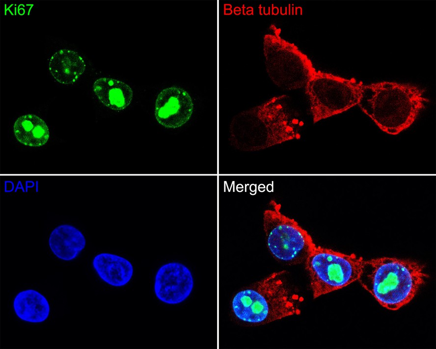
Antigen KI-67, also known as Ki-67, Ki-67 or MKI67 (Marker Of Proliferation Ki-67), is a protein that in humans is encoded by the MKI67 gene (antigen identified by monoclonal antibody Ki-67). Antigen KI-67 is a nuclear protein that is associated with cellular proliferation. Altering Ki-67 expression levels did not significantly affect cell proliferation in vivo. Ki-67 mutant mice developed normally and cells lacking Ki-67 proliferated efficiently. Furthermore, it is associated with ribosomal RNA transcription. Inactivation of antigen KI-67 leads to inhibition of ribosomal RNA synthesis. The Ki-67 protein (also known as MKI67) is a cellular marker for proliferation, and can be used in immunohistochemistry. It is strictly associated with cell proliferation. During interphase, the Ki-67 antigen can be exclusively detected within the cell nucleus, whereas in mitosis most of the protein is relocated to the surface of the chromosomes. Ki-67 protein is present during all active phases of the cell cycle (G1, S, G2, and mitosis), but is absent in resting (quiescent) cells (G0). Cellular content of Ki-67 protein markedly increases during cell progression through S phase of the cell cycle. In breast cancer Ki67 identifies a high proliferative subset of patients with ER-positive breast cancer who derive greater benefit from adjuvant chemotherapy. Ki-67 is an excellent marker to determine the growth fraction of a given cell population. The fraction of Ki-67-positive tumor cells (the Ki-67 labeling index) is often correlated with the clinical course of cancer. The best-studied examples in this context are prostate, brain and breast carcinomas, as well as nephroblastoma and neuroendocrine tumors. For these types of tumors, the prognostic value for survival and tumor recurrence have repeatedly been proven in uni- and multivariate analysis.
Background References
1. Zhang A et al. The Role of Ki67 in Evaluating Neoadjuvant Endocrine Therapy of Hormone Receptor-Positive Breast Cancer. Front Endocrinol (Lausanne). 2021 Nov
2. Travaglino A et al. Ki67 as a prognostic marker in uterine leiomyosarcoma: A quantitative systematic review. Eur J Obstet Gynecol Reprod Biol. 2021 Nov
Subcellular Location
Chromosome, Nucleus, nucleolus.
UNIPROT
Synonyms
Antigen identified by monoclonal antibody Ki 67 antibody
Antigen identified by monoclonal antibody Ki-67 antibody
Antigen KI-67 antibody
Antigen KI67 antibody
Antigen Ki67 antibody
KI67_HUMAN antibody
KIA antibody
Marker of proliferation Ki-67 antibody
MIB 1 antibody
MIB antibody
ExpandAntigen identified by monoclonal antibody Ki 67 antibody
Antigen identified by monoclonal antibody Ki-67 antibody
Antigen KI-67 antibody
Antigen KI67 antibody
Antigen Ki67 antibody
KI67_HUMAN antibody
KIA antibody
Marker of proliferation Ki-67 antibody
MIB 1 antibody
MIB antibody
MKI67 antibody
PPP1R105 antibody
Proliferation marker protein Ki-67 antibody
Proliferation related Ki 67 antigen antibody
Protein phosphatase 1 regulatory subunit 105 antibody
RP11-380J17.2 antibody
CollapseImages
-
Immunohistochemical analysis of paraffin-embedded human cervix carcinoma tissue with Rabbit anti-Ki67 antibody (HA721254) at 1/2,000 dilution.
The section was pre-treated using heat mediated antigen retrieval with sodium citrate buffer (pH 6.0) for 2 minutes. The tissues were blocked in 1% BSA for 20 minutes at room temperature, washed with ddH2O and PBS, and then probed with the primary antibody (HA721254) at 1/2,000 dilution for 1 hour at room temperature. The detection was performed using an HRP conjugated compact polymer system. DAB was used as the chromogen. Tissues were counterstained with hematoxylin and mounted with DPX. -
Immunohistochemical analysis of paraffin-embedded human colon carcinoma tissue with Rabbit anti-Ki67 antibody (HA721254) at 1/2,000 dilution.
The section was pre-treated using heat mediated antigen retrieval with sodium citrate buffer (pH 6.0) for 2 minutes. The tissues were blocked in 1% BSA for 20 minutes at room temperature, washed with ddH2O and PBS, and then probed with the primary antibody (HA721254) at 1/2,000 dilution for 1 hour at room temperature. The detection was performed using an HRP conjugated compact polymer system. DAB was used as the chromogen. Tissues were counterstained with hematoxylin and mounted with DPX. -
Immunohistochemical analysis of paraffin-embedded human lymph nodes tissue with Rabbit anti-Ki67 antibody (HA721254) at 1/2,000 dilution.
The section was pre-treated using heat mediated antigen retrieval with sodium citrate buffer (pH 6.0) for 2 minutes. The tissues were blocked in 1% BSA for 20 minutes at room temperature, washed with ddH2O and PBS, and then probed with the primary antibody (HA721254) at 1/2,000 dilution for 1 hour at room temperature. The detection was performed using an HRP conjugated compact polymer system. DAB was used as the chromogen. Tissues were counterstained with hematoxylin and mounted with DPX. -
Flow cytometric analysis of A431 cells labeling Ki67.
Cells were fixed and permeabilized. Then stained with the primary antibody (HA721254, 1ug/ml) (red) compared with Rabbit IgG Isotype Control (green). After incubation of the primary antibody at +4℃ for an hour, the cells were stained with a iFluor™ 488 conjugate-Goat anti-Rabbit IgG Secondary antibody (HA1121) at 1/1,000 dilution for 30 minutes at +4℃. Unlabelled sample was used as a control (cells without incubation with primary antibody; black). -
Flow cytometric analysis of Hela cells labeling Ki67.
Cells were fixed and permeabilized. Then stained with the primary antibody (HA721254, 1ug/ml) (red) compared with Rabbit IgG Isotype Control (green). After incubation of the primary antibody at +4℃ for an hour, the cells were stained with a iFluor™ 488 conjugate-Goat anti-Rabbit IgG Secondary antibody (HA1121) at 1/1,000 dilution for 30 minutes at +4℃. Unlabelled sample was used as a control (cells without incubation with primary antibody; black).
Please note: All products are "FOR RESEARCH USE ONLY AND ARE NOT INTENDED FOR DIAGNOSTIC OR THERAPEUTIC USE"
Products with the same target and pathway
Ki67 Rabbit Polyclonal Antibody
Application: WB,IHC-P,IF-Cell
Reactivity: Human
Conjugate: unconjugated
Ki67 Recombinant Rabbit Monoclonal Antibody [PSH0-02] - BSA and Azide free
Application: IHC-P,FC
Reactivity: Human
Conjugate: unconjugated
iFluor™ 647 Conjugated Ki67 Recombinant Rabbit Monoclonal Antibody [SR00-02]
Application: IF-Cell,IF-Tissue,FC,IHC-Fr
Reactivity: Human,Mouse
Conjugate: iFluor™ 647
Ki67 Rabbit Polyclonal Antibody
Application: WB,IF-Cell,IHC-P,FC
Reactivity: Human
Conjugate: unconjugated
Ki67 Recombinant Mouse Monoclonal Antibody [PD00-10]
Application: IHC-P,IF-Tissue
Reactivity: Human,Mouse
Conjugate: unconjugated
Ki67 Recombinant Rabbit Monoclonal Antibody [ST50-01]
Application: WB,IF-Cell,IHC-P,FC
Reactivity: Human
Conjugate: unconjugated
Ki67 Rabbit Polyclonal Antibody
Application: WB,IP,IF,IHC-P
Reactivity: Human
Conjugate: unconjugated
Ki67 Recombinant Rabbit Monoclonal Antibody [SR00-02]
Application: IHC-P,IF-Tissue,mIHC,IF-Cell,FC,IHC-Fr
Reactivity: Human,Mouse,Rat,Cynomolgus monkey
Conjugate: unconjugated
Ki67 Recombinant Rabbit Monoclonal Antibody
Application: mIHC
Reactivity: Human
Conjugate: unconjugated
iFluor™ 488 Conjugated Ki67 Recombinant Rabbit Monoclonal Antibody [SR00-02]
Application: IF-Cell,IF-Tissue,FC
Reactivity: Human
Conjugate: iFluor™ 488