Beta Catenin Recombinant Rabbit Monoclonal Antibody [SA30-04] - BSA and Azide free
Specification
Catalog# HA750023
Beta Catenin Recombinant Rabbit Monoclonal Antibody [SA30-04] - BSA and Azide free
-
WB
-
IHC-P
-
IF-Tissue
-
IP
-
IF-Cell
-
IHC-Fr
-
FC
-
Human
-
Mouse
-
Rat
-
ET1601-5
含抗保成分
-
unconjugated
Overview
Product Name
Beta Catenin Recombinant Rabbit Monoclonal Antibody [SA30-04] - BSA and Azide free
Antibody Type
Recombinant Rabbit monoclonal Antibody
Immunogen
Synthetic peptide within human Beta-Catenin aa 30-70.
Species Reactivity
Human, Mouse, Rat
Validated Applications
WB, IHC-P, IF-Tissue, IP, IF-Cell, IHC-Fr, FC
Molecular Weight
Predicted band size: 85 kDa
Positive Control
SW480 cell lysate, A431 cell lysate, HT-29 cell lysates, NIH/3T3 cell lysate, rat brain tissue lysate, mouse pancreas, mouse liver, human colon cancer tissue, mouse colon tissue, A431, C6.
Conjugation
unconjugated
Clone Number
SA30-04
Product Features
Form
Liquid
Storage Instructions
Store at +4℃ after thawing. Aliquot store at -20℃ or -80℃. Avoid repeated freeze / thaw cycles.
Storage Buffer
PBS (pH7.4).
Isotype
IgG
Purification Method
Protein A affinity purified.
Application Dilution
-
WB
-
1:1,000-1:2,000
-
IHC-P
-
1:200-1:1,000
-
IF-Tissue
-
1:100
-
IP
-
1-2μg/sample
-
IF-Cell
-
1:100
-
IHC-Fr
-
1:200
-
FC
-
1:1,000
Applications in Publications
Species in Publications
Target
Function
Catenin beta-1, also known as beta-catenin (β-catenin), is a protein that in humans is encoded by the CTNNB1 gene. Beta-catenin is a dual function protein, involved in regulation and coordination of cell–cell adhesion and gene transcription. In humans, the CTNNB1 protein is encoded by the CTNNB1 gene. In Drosophila, the homologous protein is called armadillo. β-catenin is a subunit of the cadherin protein complex and acts as an intracellular signal transducer in the Wnt signaling pathway. Mutations and overexpression of β-catenin are associated with many cancers, including hepatocellular carcinoma, colorectal carcinoma, lung cancer, malignant breast tumors, ovarian and endometrial cancer. Alterations in the localization and expression levels of beta-catenin have been associated with various forms of heart disease, including dilated cardiomyopathy. β-catenin is regulated and destroyed by the beta-catenin destruction complex, and in particular by the adenomatous polyposis coli (APC) protein, encoded by the tumour-suppressing APC gene. Therefore, genetic mutation of the APC gene is also strongly linked to cancers, and in particular colorectal cancer resulting from familial adenomatous polyposis (FAP).
Background References
1. "AlphaT-catenin: a novel tissue-specific beta-catenin-binding protein mediating strong cell-cell adhesion."Janssens B., Goossens S., Staes K., Gilbert B., van Hengel J., Colpaert C., Bruyneel E., Mareel M., van Roy F. J. Cell Sci. 114:3177-3188(2001).
2. "Characterisation of the phosphorylation of beta-catenin at the GSK-3 priming site Ser45." Hagen T., Vidal-Puig A. Biochem. Biophys. Res. Commun. 294:324-328(2002).
Sequence Similarity
Belongs to the beta-catenin family.
Tissue Specificity
Expressed in several hair follicle cell types: basal and peripheral matrix cells, and cells of the outer and inner root sheaths. Expressed in colon. Present in cortical neurons (at protein level). Expressed in breast cancer tissues (at protein level).
Post-translational Modification
Phosphorylation at Ser-552 by AMPK promotes stabilizion of the protein, enhancing TCF/LEF-mediated transcription (By similarity). Phosphorylation by GSK3B requires prior phosphorylation of Ser-45 by another kinase. Phosphorylation proceeds then from Thr-41 to Ser-37 and Ser-33. Phosphorylated by NEK2. EGF stimulates tyrosine phosphorylation. Phosphorylation on Tyr-654 decreases CDH1 binding and enhances TBP binding. Phosphorylated on Ser-33 and Ser-37 by HIPK2 and GSK3B, this phosphorylation triggers proteasomal degradation. Phosphorylation on Ser-191 and Ser-246 by CDK5. Phosphorylation by CDK2 regulates insulin internalization. Phosphorylation by PTK6 at Tyr-64, Tyr-142, Tyr-331 and/or Tyr-333 with the predominant site at Tyr-64 is not essential for inhibition of transcriptional activity.; Ubiquitinated by the SCF(BTRC) E3 ligase complex when phosphorylated by GSK3B, leading to its degradation. Ubiquitinated by a E3 ubiquitin ligase complex containing UBE2D1, SIAH1, CACYBP/SIP, SKP1, APC and TBL1X, leading to its subsequent proteasomal degradation. Ubiquitinated and degraded following interaction with SOX9 (By similarity).; S-nitrosylation at Cys-619 within adherens junctions promotes VEGF-induced, NO-dependent endothelial cell permeability by disrupting interaction with E-cadherin, thus mediating disassembly adherens junctions.; O-glycosylation at Ser-23 decreases nuclear localization and transcriptional activity, and increases localization to the plasma membrane and interaction with E-cadherin CDH1.; Deacetylated at Lys-49 by SIRT1.
Subcellular Location
Cytoplasm, Nucleus, Cell membrane, Cell junction
Synonyms
β Catenin
Beta catenin antibody
Beta-catenin antibody
Cadherin associated protein antibody
Catenin (cadherin associated protein), beta 1, 88kDa antibody
Catenin beta 1 antibody
Catenin beta-1 antibody
CATNB antibody
CHBCAT antibody
CTNB1_HUMAN antibody
Expandβ Catenin
Beta catenin antibody
Beta-catenin antibody
Cadherin associated protein antibody
Catenin (cadherin associated protein), beta 1, 88kDa antibody
Catenin beta 1 antibody
Catenin beta-1 antibody
CATNB antibody
CHBCAT antibody
CTNB1_HUMAN antibody
CTNNB antibody
CTNNB1 antibody
DKFZp686D02253 antibody
FLJ25606 antibody
FLJ37923 antibody
OTTHUMP00000162082 antibody
OTTHUMP00000165222 antibody
OTTHUMP00000165223 antibody
OTTHUMP00000209288 antibody
OTTHUMP00000209289 antibody
CollapseImages
-

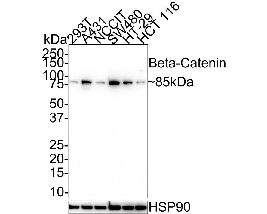
Western blot analysis of Beta Catenin on different lysates with Rabbit anti-Beta Catenin antibody (HA750023) at 1/2,000 dilution.
Lane 1: SW480 cell lysate
Lane 2: A431 cell lysate
Lane 3: HT-29 cell lysate
Lysates/proteins at 20 µg/Lane.
Predicted band size: 85 kDa
Observed band size: 100 kDa
Exposure time: 3 minutes;
4-20% SDS-PAGE gel.
Proteins were transferred to a PVDF membrane and blocked with 5% NFDM/TBST for 1 hour at room temperature. The primary antibody (HA750023) at 1/2,000 dilution was used in 5% NFDM/TBST at 4℃ overnight. Goat Anti-Rabbit IgG - HRP Secondary Antibody (HA1001) at 1/50,000 dilution was used for 1 hour at room temperature. -

☑ Knockdown (KD)
Western blot analysis of Beta Catenin on different lysates with Rabbit anti-Beta Catenin antibody (HA750023) at 1/2,000 dilution.
Lane 1: THP-1-si NT cell lysate
Lane 2: THP-1-si Beta Catenin cell lysate
Lysates/proteins at 10 µg/Lane.
Predicted band size: 85 kDa
Observed band size: 85 kDa
Exposure time: 3 minutes; ECL: K1801;
4-20% SDS-PAGE gel.
Proteins were transferred to a PVDF membrane and blocked with 5% NFDM/TBST for 1 hour at room temperature. The primary antibody (HA750023) at 1/2,000 dilution was used in primary antibody dilution at 4℃ overnight. Goat Anti-Rabbit IgG - HRP Secondary Antibody (HA1001) at 1/50,000 dilution was used for 1 hour at room temperature. -

☑ Knockdown (KD)
Western blot analysis of Beta Catenin on different lysates with Rabbit anti-Beta Catenin antibody (HA750023) at 1/2,000 dilution.
Lane 1: HAP1-parental cell lysate
Lane 2: HAP1-Beta Catenin KD cell lysate
Lysates/proteins at 10 µg/Lane.
Predicted band size: 85 kDa
Observed band size: 85 kDa
Exposure time: 40 seconds; ECL: K1801;
4-20% SDS-PAGE gel.
Proteins were transferred to a PVDF membrane and blocked with 5% NFDM/TBST for 1 hour at room temperature. The primary antibody (HA750023) at 1/2,000 dilution was used in K1803 at 4℃ overnight. Goat Anti-Rabbit IgG - HRP Secondary Antibody (HA1001) at 1/50,000 dilution was used for 1 hour at room temperature. -

Western blot analysis of Beta Catenin on different lysates with Rabbit anti-Beta Catenin antibody (HA750023) at 1/1,000 dilution.
Lane 1: NIH/3T3 cell lysate (10 µg/Lane)
Lane 2: Rat brain tissue lysate (20 µg/Lane)
Predicted band size: 85 kDa
Observed band size: 85 kDa
Exposure time: 30 seconds;
6% SDS-PAGE gel.
Proteins were transferred to a PVDF membrane and blocked with 5% NFDM/TBST for 1 hour at room temperature. The primary antibody (HA750023) at 1/1,000 dilution was used in 5% NFDM/TBST at room temperature for 2 hours. Goat Anti-Rabbit IgG - HRP Secondary Antibody (HA1001) at 1:100,000 dilution was used for 1 hour at room temperature. -

Immunohistochemical analysis of paraffin-embedded human colon cancer tissue with Rabbit anti-Beta Catenin antibody (HA750023) at 1/1,000 dilution.
The section was pre-treated using heat mediated antigen retrieval with sodium citrate buffer (pH 6.0) (high pressure) for 2 minutes. The tissues were blocked in 1% BSA for 20 minutes at room temperature, washed with ddH2O and PBS, and then probed with the primary antibody (HA750023) at 1/1,000 dilution for 1 hour at room temperature. The detection was performed using an HRP conjugated compact polymer system. DAB was used as the chromogen. Tissues were counterstained with hematoxylin and mounted with DPX. -

Immunohistochemical analysis of paraffin-embedded mouse colon tissue with Rabbit anti-Beta Catenin antibody (HA750023) at 1/1,000 dilution.
The section was pre-treated using heat mediated antigen retrieval with sodium citrate buffer (pH 6.0) (high pressure) for 2 minutes. The tissues were blocked in 1% BSA for 20 minutes at room temperature, washed with ddH2O and PBS, and then probed with the primary antibody (HA750023) at 1/1,000 dilution for 1 hour at room temperature. The detection was performed using an HRP conjugated compact polymer system. DAB was used as the chromogen. Tissues were counterstained with hematoxylin and mounted with DPX. -

Immunohistochemical analysis of paraffin-embedded rat brain tissue with Rabbit anti-Beta Catenin antibody (HA750023) at 1/200 dilution.
The section was pre-treated using heat mediated antigen retrieval with sodium citrate buffer (pH 6.0) (high pressure) for 2 minutes. The tissues were blocked in 1% BSA for 20 minutes at room temperature, washed with ddH2O and PBS, and then probed with the primary antibody (HA750023) at 1/200 dilution for 1 hour at room temperature. The detection was performed using an HRP conjugated compact polymer system. DAB was used as the chromogen. Tissues were counterstained with hematoxylin and mounted with DPX. -

Immunofluorescence analysis of paraffin-embedded human colon cancer tissue labeling Beta Catenin with Rabbit anti-Beta Catenin antibody (HA750023) at 1/100 dilution.
The section was pre-treated using heat mediated antigen retrieval with Tris-EDTA buffer (pH 9.0) for 20 minutes. The tissues were blocked in 10% negative goat serum for 1 hour at room temperature, washed with PBS, and then probed with the primary antibody (HA750023, green) at 1/100 dilution overnight at 4 ℃, washed with PBS. Goat Anti-Rabbit IgG H&L (iFluor™ 488, HA1121) was used as the secondary antibody at 1/1,000 dilution. Nuclei were counterstained with DAPI (blue). -

Immunofluorescence analysis of paraffin-embedded mouse colon tissue labeling Beta Catenin with Rabbit anti-Beta Catenin antibody (HA750023) at 1/100 dilution.
The section was pre-treated using heat mediated antigen retrieval with Tris-EDTA buffer (pH 9.0) for 20 minutes. The tissues were blocked in 10% negative goat serum for 1 hour at room temperature, washed with PBS, and then probed with the primary antibody (HA750023, green) at 1/100 dilution overnight at 4 ℃, washed with PBS. Goat Anti-Rabbit IgG H&L (iFluor™ 488, HA1121) was used as the secondary antibody at 1/1,000 dilution. Nuclei were counterstained with DAPI (blue). -

Immunocytochemistry analysis of A431 cells labeling Beta Catenin with Rabbit anti-Beta Catenin antibody (HA750023) at 1/100 dilution.
Cells were fixed in 4% paraformaldehyde for 20 minutes at room temperature, permeabilized with 0.1% Triton X-100 in PBS for 5 minutes at room temperature, then blocked with 1% BSA in 10% negative goat serum for 1 hour at room temperature. Cells were then incubated with Rabbit anti-Beta Catenin antibody (HA750023) at 1/100 dilution in 1% BSA in PBST overnight at 4 ℃. Goat Anti-Rabbit IgG H&L (iFluor™ 488, HA1121) was used as the secondary antibody at 1/1,000 dilution. PBS instead of the primary antibody was used as the secondary antibody only control. Nuclear DNA was labelled in blue with DAPI. Beta tubulin (M1305-2, red) was stained at 1/100 dilution overnight at +4℃. Goat Anti-Mouse IgG H&L (iFluor™ 594, HA1126) was used as the secondary antibody at 1/1,000 dilution. -

Immunocytochemistry analysis of C6 cells labeling Beta Catenin with Rabbit anti-Beta Catenin antibody (HA750023) at 1/100 dilution.
Cells were fixed in 4% paraformaldehyde for 20 minutes at room temperature, permeabilized with 0.1% Triton X-100 in PBS for 5 minutes at room temperature, then blocked with 1% BSA in 10% negative goat serum for 1 hour at room temperature. Cells were then incubated with Rabbit anti-Beta Catenin antibody (HA750023) at 1/100 dilution in 1% BSA in PBST overnight at 4 ℃. Goat Anti-Rabbit IgG H&L (iFluor™ 488, HA1121) was used as the secondary antibody at 1/1,000 dilution. PBS instead of the primary antibody was used as the secondary antibody only control. Nuclear DNA was labelled in blue with DAPI. Beta tubulin (M1305-2, red) was stained at 1/100 dilution overnight at +4℃. Goat Anti-Mouse IgG H&L (iFluor™ 594, HA1126) was used as the secondary antibody at 1/1,000 dilution. -

Immunofluorescence analysis of frozen mouse colon tissue with Rabbit anti-Beta Catenin antibody (HA750023) at 1/200 dilution.
The section was pre-treated using heat mediated antigen retrieval with sodium citrate buffer (pH 6.0) for about 2 minutes in microwave oven. The tissues were blocked in 10% negative goat serum for 1 hour at room temperature, washed with PBS, and then probed with the primary antibody (HA750023, green) at 1/200 dilution overnight at 4 ℃, washed with PBS. Goat Anti-Rabbit IgG H&L (iFluor™ 488, HA1121) was used as the secondary antibody at 1/1,000 dilution. Nuclei were counterstained with DAPI (blue). -

Beta Catenin was immunoprecipitated from 0.2 mg rat brain tissue lysate with HA750023 at 2 µg/10 µl beads. Western blot was performed from the immunoprecipitate using HA750023 at 1/1,000 dilution. Anti-Rabbit IgG for IP Nano-secondary antibody (NBI01H) at 1/5,000 dilution was used for 1 hour at room temperature.
Lane 1: Rat brain tissue lysate (input)
Lane 2: HA750023 IP in rat brain tissue lysate
Lane 3: Rabbit IgG instead of HA750023 in rat brain tissue lysate
Blocking/Dilution buffer: 5% NFDM/TBST
Exposure time: 2 seconds; ECL: K1801 -

Flow cytometric analysis of A431 cells labeling Beta Catenin.
Cells were fixed and permeabilized. Then stained with the primary antibody (HA750023, 1/1,000) (red) compared with Rabbit IgG Isotype Control (green). After incubation of the primary antibody at +4℃ for an hour, the cells were stained with a iFluor™ 488 conjugate-Goat anti-Rabbit IgG Secondary antibody (HA1121) at 1/1,000 dilution for 30 minutes at +4℃. Unlabelled sample was used as a control (cells without incubation with primary antibody; black).
Please note: All products are "FOR RESEARCH USE ONLY AND ARE NOT INTENDED FOR DIAGNOSTIC OR THERAPEUTIC USE"
Citation
-
Adhesive Hydrogel Barriers Synergistically Promote Bone Regeneration by Self-Constructing Microstress and Mineralization Microenvironment
Author: Senlin Chen, Mingxin Qiao, Yanhua Liu, Zihan He, Shihua Huang, Zhengyi Xu, Wenjia Xie, Jian Wang, Zhou Zhu, Qianbing Wan
PMID: 40421766
Journal: Journal Of Materials Chemistry B
Application: IF
Reactivity: Rat
Publish date: 2025 May
-
Citation
Products with the same target and pathway
Beta Catenin Rabbit Polyclonal Antibody
Application: WB,IF-Cell
Reactivity: Human,Mouse,Rat,Zebrafish
Conjugate: unconjugated
Beta Catenin Mouse Monoclonal Antibody [10-C0-B7]
Application: WB,IF-Cell,IHC-P,FC
Reactivity: Human,Mouse,Rat
Conjugate: unconjugated
Phospho-Beta Catenin (S552) Recombinant Rabbit Monoclonal Antibody [PSH08-72] - BSA and Azide free
Application: WB,IF-Cell,IHC-P,FC
Reactivity: Human,Mouse,Rat
Conjugate: unconjugated
Beta Catenin Recombinant Antibody - Rat IgG1 (Chimeric)
Application: mIHC
Reactivity: Human
Conjugate: unconjugated
Beta Catenin Mouse Monoclonal Antibody [A6-F8]
Application: WB,IF-Cell,IHC-P,FC,IF-Tissue,ChIP
Reactivity: Human,Mouse,Rat
Conjugate: unconjugated
Phospho-Beta Catenin (T41/S45) Recombinant Rabbit Monoclonal Antibody [JE54-02]
Application: WB,IHC-P
Reactivity: Human,Mouse,Rat
Conjugate: unconjugated
Phospho-Beta Catenin (S552) Recombinant Rabbit Monoclonal Antibody [PSH08-72]
Application: WB,IF-Cell,IHC-P,FC
Reactivity: Human,Mouse,Rat
Conjugate: unconjugated
Beta Catenin Recombinant Mouse Monoclonal Antibody [A6-F8-R] - BSA and Azide free
Application: WB,IF-Cell,FC
Reactivity: Human,Mouse,Rat
Conjugate: unconjugated
Beta Catenin Recombinant Rabbit Monoclonal Antibody [SA30-04]
Application: WB,IHC-P,IF-Tissue,IP,mIHC,IF-Cell,IHC-Fr,FC
Reactivity: Human,Mouse,Rat
Conjugate: unconjugated
Beta Catenin Recombinant Antibody - Mouse IgG1 (Chimeric)
Application: mIHC
Reactivity: Human
Conjugate: unconjugated
Beta Catenin Rabbit Polyclonal Antibody
Application: WB,IHC-P,FC,IF-Cell,IF-Tissue
Reactivity: Human,Mouse,Rat
Conjugate: unconjugated
Phospho-Beta Catenin (S33 + S37) Recombinant Rabbit Monoclonal Antibody [JE59-59]
Application: WB
Reactivity: Human,Rat,Mouse
Conjugate: unconjugated
iFluor™ 488 Conjugated Beta Catenin Recombinant Rabbit Monoclonal Antibody [SA30-04]
Application: IF-Tissue,IF-Cell
Reactivity: Human,Mouse
Conjugate: iFluor™ 488
Beta Catenin Recombinant Mouse Monoclonal Antibody [A6-F8-R]
Application: WB,IF-Cell,FC
Reactivity: Human,Mouse,Rat
Conjugate: unconjugated
Beta Catenin Recombinant Rabbit Monoclonal Antibody
Application: mIHC
Reactivity: Human
Conjugate: unconjugated


