E-Cadherin Rabbit Polyclonal Antibody
Specification
Catalog# 0407-25
E-Cadherin Rabbit Polyclonal Antibody
-
WB
-
IF-Cell
-
FC
-
Human
-
Mouse
-
unconjugated
Overview
Product Name
E-Cadherin Rabbit Polyclonal Antibody
Antibody Type
Rabbit Polyclonal Antibody
Immunogen
Synthetic peptide within mouse E-Cadherin aa 595-644 / 882.
Species Reactivity
Human (Predicted: Mouse)
Validated Applications
WB, IF-Cell, FC
Molecular Weight
Predicted band size: 97 kDa
Positive Control
HCT116, SKOV-3, Hela.
Conjugation
unconjugated
RRID
Product Features
Form
Liquid
Storage Instructions
Shipped at 4℃. Store at +4℃ short term (1-2 weeks). It is recommended to aliquot into single-use upon delivery. Store at -20℃ long term.
Storage Buffer
1*PBS (pH7.4), 0.2% BSA, 40% Glycerol. Preservative: 0.05% Sodium Azide.
Isotype
IgG
Purification Method
Immunogen affinity purified.
Application Dilution
-
WB
-
1:500
-
IF-Cell
-
1:50-1:200
-
FC
-
1:50-1:100
Applications in Publications
Species in Publications
| Human | See 7 publications below |
| Mouse | See 4 publications below |
Target
Function
Cadherin-1 or Epithelial cadherin (E-cadherin), (not to be confused with the APC/C activator protein CDH1) is a protein that in humans is encoded by the CDH1 gene. Mutations are correlated with gastric, breast, colorectal, thyroid, and ovarian cancers. CDH1 has also been designated as CD324 (cluster of differentiation 324). It is a tumor suppressor gene. Cadherin-1 is a classical member of the cadherin superfamily. The encoded protein is a calcium-dependent cell–cell adhesion glycoprotein composed of five extracellular cadherin repeats, a transmembrane region, and a highly conserved cytoplasmic tail. Mutations in this gene are correlated with gastric, breast, colorectal, thyroid, and ovarian cancers. Loss of function is thought to contribute to progression in cancer by increasing proliferation, invasion, and/or metastasis. The ectodomain of this protein mediates bacterial adhesion to mammalian cells, and the cytoplasmic domain is required for internalization. Identified transcript variants arise from mutation at consensus splice sites. E-cadherin (epithelial) is the most well-studied member of the cadherin family. It consists of 5 cadherin repeats (EC1 ~ EC5) in the extracellular domain, one transmembrane domain, and an intracellular domain that binds p120-catenin and beta-catenin. The intracellular domain contains a highly-phosphorylated region vital to beta-catenin binding and, therefore, to E-cadherin function.
Background References
1. Meigs T.E et al. Galpha12 and Galpha13 negatively regulate the adhesive functions of cadherin. J Biol Chem 277:24594-24600 (2002).
2. Agiostratidou G et al. The cytoplasmic sequence of E-cadherin promotes non-amyloidogenic degradation of A beta precursors. J Neurochem 96:1182-1188 (2006).
Tissue Specificity
Non-neural epithelial tissues.
Post-translational Modification
During apoptosis or with calcium influx, cleaved by a membrane-bound metalloproteinase (ADAM10), PS1/gamma-secretase and caspase-3. Processing by the metalloproteinase, induced by calcium influx, causes disruption of cell-cell adhesion and the subsequent release of beta-catenin into the cytoplasm. The residual membrane-tethered cleavage product is rapidly degraded via an intracellular proteolytic pathway. Cleavage by caspase-3 releases the cytoplasmic tail resulting in disintegration of the actin microfilament system. The gamma-secretase-mediated cleavage promotes disassembly of adherens junctions. During development of the cochlear organ of Corti, cleavage by ADAM10 at adherens junctions promotes pillar cell separation (By similarity).; N-glycosylation at Asn-637 is essential for expression, folding and trafficking. Addition of bisecting N-acetylglucosamine by MGAT3 modulates its cell membrane location.; Ubiquitinated by a SCF complex containing SKP2, which requires prior phosphorylation by CK1/CSNK1A1. Ubiquitinated by CBLL1/HAKAI, requires prior phosphorylation at Tyr-754.; O-glycosylated. O-manosylated by TMTC1, TMTC2, TMTC3 or TMTC4. Thr-285 and Thr-509 are O-mannosylated by TMTC2 or TMTC4 but not TMTC1 or TMTC3.
Subcellular Location
Cell membrane.
Synonyms
Arc 1 antibody
CADH1_HUMAN antibody
Cadherin 1 antibody
cadherin 1 type 1 E-cadherin antibody
Cadherin1 antibody
CAM 120/80 antibody
CD 324 antibody
CD324 antibody
CD324 antigen antibody
cdh1 antibody
ExpandArc 1 antibody
CADH1_HUMAN antibody
Cadherin 1 antibody
cadherin 1 type 1 E-cadherin antibody
Cadherin1 antibody
CAM 120/80 antibody
CD 324 antibody
CD324 antibody
CD324 antigen antibody
cdh1 antibody
CDHE antibody
E-Cad/CTF3 antibody
E Cadherin antibody
E-cadherin antibody
ECAD antibody
Epithelial cadherin antibody
epithelial calcium dependant adhesion protein antibody
LCAM antibody
Liver cell adhesion molecule antibody
UVO antibody
Uvomorulin antibody
CollapseImages
-

Western blot analysis of E-Cadherin on A431 cell lysate. Proteins were transferred to a PVDF membrane and blocked with 5% BSA in PBS for 1 hour at room temperature. The primary antibody was used at a 1:500 dilution in 5% BSA at room temperature for 2 hours. Goat Anti-Rabbit IgG - HRP Secondary Antibody (HA1001) at 1:5,000 dilution was used for 1 hour at room temperature.
-

ICC staining E-Cadherin in HCT116 cells (green). Formalin fixed cells were permeabilized with 0.1% Triton X-100 in TBS for 10 minutes at room temperature and blocked with 1% Blocker BSA for 15 minutes at room temperature. Cells were probed with the antibody (0407-25) at a dilution of 1:200 for 1 hour at room temperature, washed with PBS. Alexa Fluorc™ 488 Goat anti-Rabbit IgG was used as the secondary antibody at 1/100 dilution.
-

ICC staining E-Cadherin in SKOV-3 cells (green). Formalin fixed cells were permeabilized with 0.1% Triton X-100 in TBS for 10 minutes at room temperature and blocked with 1% Blocker BSA for 15 minutes at room temperature. Cells were probed with the antibody (0407-25) at a dilution of 1:200 for 1 hour at room temperature, washed with PBS. Alexa Fluorc™ 488 Goat anti-Rabbit IgG was used as the secondary antibody at 1/100 dilution.
-

Flow cytometric analysis of E-Cadherin was done on Hela cells. The cells were fixed, permeabilized and stained with E-Cadherin antibody at 1/100 dilution (blue) compared with an unlabelled control (cells without incubation with primary antibody; red). After incubation of the primary antibody on room temperature for an hour, the cells was stained with a Alexa Fluor™ 488-conjugated goat anti-rabbit IgG Secondary antibody at 1/500 dilution for 30 minutes.
Please note: All products are "FOR RESEARCH USE ONLY AND ARE NOT INTENDED FOR DIAGNOSTIC OR THERAPEUTIC USE"
Citation
-
Integrated multi-omics elucidates PRNP knockdown-mediated chemosensitization to gemcitabine in pancreatic ductal adenocarcinoma
Author: Bing Qi, Yuwei Wu, Ning Wang, Liumeng Duan, Yangbo Wei, Xuanrui Zhang, Jing Chen
PMID: 10.3389/fimmu.2025.1667835
Journal: Frontiers In Immunology
Application: WB
Reactivity: Human
Publish date: 2025 Nov
-
Citation
-
The RNA-binding protein DDX39B promotes colorectal adenocarcinoma progression by stabilizing DCLK1
Author:
PMID: 40583564
Journal: HUMAN MOLECULAR GENETICS
Application: WB
Reactivity: Human
Publish date: 2025 Jun
-
Citation
-
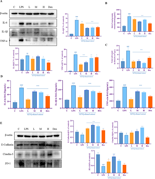
Sanzi Yangqin Decoction improved acute lung injury by regulating the TLR2-mediated NF-κB/NLRP3 signaling pathway and inhibiting the activation of NLRP3 inflammasome
Author: Anna Gan, Haimiao Chen, Fei Lin, Ruixuan Wang, Bo Wu, Tingxu Yan, Ying Jia
PMID: 10.1016/j.phymed.2025.156438
Journal: Phytomedicine
Application: WB
Reactivity: Mouse
Publish date: 2025 Jan
-
Citation
-
Sanzi Yangqin Decoction improved acute lung injury by regulating the TLR2-mediated NF-κB/NLRP3 signaling pathway and inhibiting the activation of NLRP3 inflammasome
Author: Anna Gan, Haimiao Chen, Fei Lin, Ruixuan Wang, Bo Wu, Tingxu Yan, Ying Jia
PMID: 39914066

Journal: Phytomedicine
Application: WB
Reactivity: Mouse
Publish date: 2025 Jan
-
Citation
-
Reduced Proline-Rich Tyrosine Kinase 2 Promotes Tumor Metastasis by Activating Epithelial–Mesenchymal Transition in Colorectal Cancer
Author: Fangquan Wu ,et al
PMID: 39414740
Journal: Digestive Diseases And Sciences
Application: WB
Reactivity: Human
Publish date: 2024 Oct
-
Citation
-
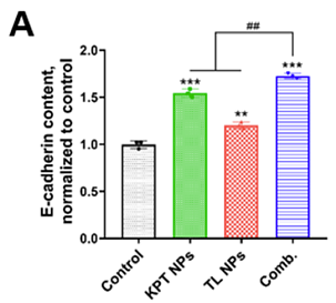
Enhance Mitochondrial Damage by Nuclear Export Inhibition to Suppress Tumor Growth and Metastasis with Increased Antitumor Properties of Macrophages
Author:
PMID: 37079389

Journal: ACS Applied Materials & Interfaces
Application: FC
Reactivity: Mouse
Publish date: 2023 May
-
Citation
-
CXCR4 inhibition attenuates calcium oxalate crystal deposition-induced renal fibrosis
Author:
PMID: 35255299

Journal: International Immunopharmacology
Application: WB
Reactivity: Mouse
Publish date: 2022 Jun
-
Citation
-
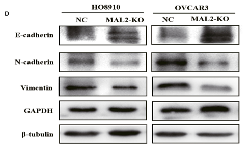
Multi-Omics Analysis of the Therapeutic Value of MAL2 Based on Data Mining in Human Cancers
Author: Yuan, J., Jiang, X., Lan, H., Zhang, X., Ding, T., Yang, F., Zeng, D., Yong, J., Niu, B., & Xiao, S.
PMID: 35111745

Journal: Frontiers In Cell And Developmental Biology
Application: WB
Reactivity: Human
Publish date: 2022 Jan
-
Citation
-
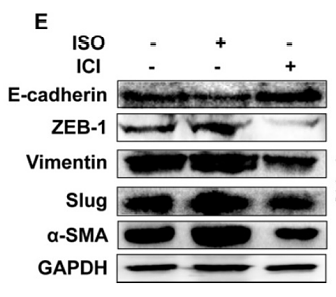
PlexinA1 activation induced by β2-AR promotes epithelial-mesenchymal transition through JAK-STAT3 signaling in human gastric cancer cells
Author: Liu, Y., Hao, Y., Zhao, H., Zhang, Y., Cheng, D., Zhao, L., Peng, Y., Lu, Y., & Li, Y.
PMID: 35517411

Journal: Journal Of Cancer
Application: WB
Reactivity: Human
Publish date: 2022 Apr
-
Citation
-
miR-888 in MCF-7 Side Population Sphere Cells Directly Targets E-cadherin
Author: Chen L
PMID: 24480745
Journal: Journal Of Genetics And Genomics
Application: WB
Reactivity: Human
Publish date: 2014 Jan
-
Citation
-
Ionizing radiation promotes migration and invasion of cancer cells through transforming growth factor-beta-mediated epithelial-mesenchymal transition.
Author: Guo GZ.
PMID: 22115555
Journal: International Journal Of Radiation Oncology, Biology, And Physics
Application: WB
Reactivity: Human
Publish date: 2011 Dec
-
Citation
Alternative Products
E-Cadherin Recombinant Rabbit Monoclonal Antibody [SY0287]
Application: WB,IF-Cell,IF-Tissue,IHC-P,FC,IP,mIHC
Reactivity: Human,Cynomolgus monkey
Conjugate: unconjugated
Products with the same target and pathway
iFluor™ 488 Conjugated E-Cadherin Recombinant Rabbit Monoclonal Antibody [SY0287]
Application: IF-Cell,FC
Reactivity: Human
Conjugate: iFluor™ 488
E-Cadherin Mouse Monoclonal Antibody [A0-G11-2]
Application: WB,IHC-P,IF-Tissue
Reactivity: Human,Mouse,Rat
Conjugate: unconjugated
E-Cadherin Recombinant Rabbit Monoclonal Antibody [PSH13-75] - BSA and Azide free
Application: WB,IHC-Fr,IHC-P,IF-Cell,FC,IP
Reactivity: Human,Mouse,Rat
Conjugate: unconjugated
E-Cadherin Recombinant Rabbit Monoclonal Antibody [SY0287] - BSA and Azide free
Application: WB,IF-Cell,IF-Tissue,IHC-P,FC,IP
Reactivity: Human,Cynomolgus monkey
Conjugate: unconjugated
iFluor™ 594 Conjugated E-Cadherin Recombinant Rabbit Monoclonal Antibody [SY0287]
Application: IF-Tissue
Reactivity: Human
Conjugate: iFluor™ 594
E-Cadherin Recombinant Mouse Monoclonal Antibody
Application: mIHC
Reactivity: Human
Conjugate: unconjugated
E-Cadherin Recombinant Rabbit Monoclonal Antibody [PSH13-75]
Application: WB,IHC-Fr,IHC-P,IF-Cell,FC,IP
Reactivity: Human,Mouse,Rat
Conjugate: unconjugated
E-Cadherin Recombinant Rabbit Monoclonal Antibody [SY0287]
Application: WB,IF-Cell,IF-Tissue,IHC-P,FC,IP,mIHC
Reactivity: Human,Cynomolgus monkey
Conjugate: unconjugated
E-Cadherin Recombinant Mouse Monoclonal Antibody [A0-G11-2-R] - BSA and Azide free
Application: WB,IHC-P
Reactivity: Human,Mouse,Rat
Conjugate: unconjugated
E-Cadherin Recombinant Mouse Monoclonal Antibody [A0-G11-2-R]
Application: WB,IHC-P,mIHC
Reactivity: Human,Mouse,Rat
Conjugate: unconjugated
E-Cadherin Rabbit Polyclonal Antibody
Application: WB,IHC-P,IF-Cell,IHC-Fr,FC
Reactivity: Human,Mouse,Rat
Conjugate: unconjugated
E-Cadherin Mouse Monoclonal Antibody [3-F9]
Application: IHC-P
Reactivity: Human,Mouse
Conjugate: unconjugated
E-Cadherin Recombinant Rabbit Monoclonal Antibody
Application: mIHC
Reactivity: Human
Conjugate: unconjugated


