E-Cadherin Rabbit Polyclonal Antibody
Specification
Catalog# ER63312
E-Cadherin Rabbit Polyclonal Antibody
-
WB
-
IHC-P
-
IF-Cell
-
IHC-Fr
-
FC
-
Human
-
Mouse
-
Rat
-
unconjugated
Overview
Product Name
E-Cadherin Rabbit Polyclonal Antibody
Antibody Type
Rabbit Polyclonal Antibody
Immunogen
Recombinant protein within mouse E-Cadherin aa 151-730.
Species Reactivity
Human, Mouse, Rat
Validated Applications
WB, IHC-P, IF-Cell, IHC-Fr, FC
Molecular Weight
Predicted band size: 97 kDa
Positive Control
T-47D cell lysate, HCT 116 cell lysate, 4T1 cell lysate, Mouse pancreas tissue lysate, Rat pancreas tissue lysate, MCF7, 4T1, mouse pancreas tissue, rat pancreas tissue, mouse colon tissue, rat colon tissue.
Conjugation
unconjugated
Product Features
Form
Liquid
Storage Instructions
Shipped at 4℃. Store at +4℃ short term (1-2 weeks). It is recommended to aliquot into single-use upon delivery. Store at -20℃ long term.
Storage Buffer
PBS (pH7.4), 0.1% BSA, 40% Glycerol. Preservative: 0.05% Sodium Azide.
Isotype
IgG
Purification Method
Immunogen affinity purified.
Application Dilution
-
WB
-
1:10,000
-
IHC-P
-
1:2,000
-
IF-Cell
-
1:500
-
IHC-Fr
-
1:1,000
-
FC
-
1:1,000
Applications in Publications
Species in Publications
| Mouse | See 3 publications below |
| Human | See 3 publications below |
Target
Function
Cadherin-1 or Epithelial cadherin (E-cadherin), (not to be confused with the APC/C activator protein CDH1) is a protein that in humans is encoded by the CDH1 gene. Mutations are correlated with gastric, breast, colorectal, thyroid, and ovarian cancers. CDH1 has also been designated as CD324 (cluster of differentiation 324). It is a tumor suppressor gene.
Background References
暂无
Subcellular Location
Cell junction, adherens junction, Cell membrane, Endosome, Golgi apparatus, trans-Golgi network, Cytoplasm, desmosome.
Synonyms
Arc 1 antibody
CADH1_HUMAN antibody
Cadherin 1 antibody
cadherin 1 type 1 E-cadherin antibody
Cadherin-1 antibody
Cadherin1 antibody
CAM 120/80 antibody
CD 324 antibody
CD324 antibody
CD324 antigen antibody
ExpandArc 1 antibody
CADH1_HUMAN antibody
Cadherin 1 antibody
cadherin 1 type 1 E-cadherin antibody
Cadherin-1 antibody
Cadherin1 antibody
CAM 120/80 antibody
CD 324 antibody
CD324 antibody
CD324 antigen antibody
cdh1 antibody
CDHE antibody
E-Cad/CTF3 antibody
E-cadherin antibody
ECAD antibody
Epithelial cadherin antibody
epithelial calcium dependant adhesion protein antibody
LCAM antibody
Liver cell adhesion molecule antibody
UVO antibody
Uvomorulin antibody
CollapseImages
-

Application: IHC-Fr
Species: Mouse
Site: Colon
Sample: Frozen section
Antibody concentration: 1/1,000
Antigen retrieval: Not required -

Application: IHC-Fr
Species: Rat
Site: Colon
Sample: Frozen section
Antibody concentration: 1/1,000
Antigen retrieval: Not required -

☑ Relative expression (RE)
Western blot analysis of E-Cadherin on different lysates with Rabbit anti-E-Cadherin antibody (ER63312) at 1/10,000 dilution.
Lane 1: T-47D cell lysate (20 µg/Lane)
Lane 2: MDA-MB-231 cell lysate (negative) (20 µg/Lane)
Lane 3: HCT 116 cell lysate (20 µg/Lane)
Lane 4: 4T1 cell lysate (20 µg/Lane)
Lane 5: C2C12 cell lysate (negative) (20 µg/Lane)
Lane 6: Mouse pancreas tissue lysate (20 µg/Lane)
Lane 7: Rat pancreas tissue lysate (20 µg/Lane)
Predicted band size: 97 kDa
Observed band size: 80-130 kDa
Exposure time: 8 seconds; ECL: K1801;
4-20% SDS-PAGE gel.
Proteins were transferred to a PVDF membrane and blocked with 5% NFDM/TBST for 1 hour at room temperature. The primary antibody (ER63312) at 1/10,000 dilution was used in 5% NFDM/TBST at 4℃ overnight. Goat Anti-Rabbit IgG - HRP Secondary Antibody (HA1001) at 1/50,000 dilution was used for 1 hour at room temperature. -

Immunocytochemistry analysis of MCF7 cells labeling E-Cadherin with Rabbit anti-E-Cadherin antibody (ER63312) at 1/500 dilution.
Cells were fixed in 4% paraformaldehyde for 15 minutes at room temperature, permeabilized with 0.1% Triton X-100 in PBS for 15 minutes at room temperature, then blocked with 1% BSA in 10% negative goat serum for 1 hour at room temperature. Cells were then incubated with Rabbit anti-E-Cadherin antibody (ER63312) at 1/500 dilution in 1% BSA in PBST overnight at 4 ℃. Goat Anti-Rabbit IgG H&L (iFluor™ 488, HA1121) was used as the secondary antibody at 1/1,000 dilution. PBS instead of the primary antibody was used as the secondary antibody only control. Nuclear DNA was labelled in blue with DAPI.
Beta tubulin (M1305-2, red) was stained at 1/100 dilution overnight at +4℃. Goat Anti-Mouse IgG H&L (iFluor™ 594, HA1126) was used as the secondary antibody at 1/1,000 dilution. -

☑ Relative expression (RE)
Immunocytochemistry analysis of 4T1 cells labeling E-Cadherin with Rabbit anti-E-Cadherin antibody (ER63312) at 1/500 dilution.
Cells were fixed in 4% paraformaldehyde for 15 minutes at room temperature, permeabilized with 0.1% Triton X-100 in PBS for 15 minutes at room temperature, then blocked with 1% BSA in 10% negative goat serum for 1 hour at room temperature. Cells were then incubated with Rabbit anti-E-Cadherin antibody (ER63312) at 1/500 dilution in 1% BSA in PBST overnight at 4 ℃. Goat Anti-Rabbit IgG H&L (iFluor™ 488, HA1121) was used as the secondary antibody at 1/1,000 dilution. PBS instead of the primary antibody was used as the secondary antibody only control. Nuclear DNA was labelled in blue with DAPI.
Beta tubulin (M1305-2, red) was stained at 1/100 dilution overnight at +4℃. Goat Anti-Mouse IgG H&L (iFluor™ 594, HA1126) was used as the secondary antibody at 1/1,000 dilution.
C2C12 is a negative control cell. -

Immunohistochemical analysis of paraffin-embedded mouse pancreas tissue with Rabbit anti-E-Cadherin antibody (ER63312) at 1/2,000 dilution.
The section was pre-treated using heat mediated antigen retrieval with Tris-EDTA buffer (pH 9.0) for 20 minutes. The tissues were blocked in 1% BSA for 20 minutes at room temperature, washed with ddH2O and PBS, and then probed with the primary antibody (ER63312) at 1/2,000 dilution for 1 hour at room temperature. The detection was performed using an HRP conjugated compact polymer system. DAB was used as the chromogen. Tissues were counterstained with hematoxylin and mounted with DPX. -

Immunohistochemical analysis of paraffin-embedded rat pancreas tissue with Rabbit anti-E-Cadherin antibody (ER63312) at 1/2,000 dilution.
The section was pre-treated using heat mediated antigen retrieval with Tris-EDTA buffer (pH 9.0) for 20 minutes. The tissues were blocked in 1% BSA for 20 minutes at room temperature, washed with ddH2O and PBS, and then probed with the primary antibody (ER63312) at 1/2,000 dilution for 1 hour at room temperature. The detection was performed using an HRP conjugated compact polymer system. DAB was used as the chromogen. Tissues were counterstained with hematoxylin and mounted with DPX. -

Immunohistochemical analysis of paraffin-embedded mouse colon tissue with Rabbit anti-E-Cadherin antibody (ER63312) at 1/2,000 dilution.
The section was pre-treated using heat mediated antigen retrieval with Tris-EDTA buffer (pH 9.0) for 20 minutes. The tissues were blocked in 1% BSA for 20 minutes at room temperature, washed with ddH2O and PBS, and then probed with the primary antibody (ER63312) at 1/2,000 dilution for 1 hour at room temperature. The detection was performed using an HRP conjugated compact polymer system. DAB was used as the chromogen. Tissues were counterstained with hematoxylin and mounted with DPX. -

Immunohistochemical analysis of paraffin-embedded rat colon tissue with Rabbit anti-E-Cadherin antibody (ER63312) at 1/2,000 dilution.
The section was pre-treated using heat mediated antigen retrieval with Tris-EDTA buffer (pH 9.0) for 20 minutes. The tissues were blocked in 1% BSA for 20 minutes at room temperature, washed with ddH2O and PBS, and then probed with the primary antibody (ER63312) at 1/2,000 dilution for 1 hour at room temperature. The detection was performed using an HRP conjugated compact polymer system. DAB was used as the chromogen. Tissues were counterstained with hematoxylin and mounted with DPX. -

Flow cytometric analysis of MCF7 cells labeling E-Cadherin.
Cells were washed twice with cold PBS and resuspend. Then stained with the primary antibody (ER63312, 1/1,000) (red) compared with Rabbit IgG Isotype Control (green). After incubation of the primary antibody at +4℃ for an hour, the cells were stained with a iFluor™ 488 conjugate-Goat anti-Rabbit IgG Secondary antibody (HA1121) at 1/1,000 dilution for 30 minutes at +4℃. Unlabelled sample was used as a control (cells without incubation with primary antibody; black). -

Flow cytometric analysis of 4T1 cells labeling E-Cadherin.
Cells were washed twice with cold PBS and resuspend. Then stained with the primary antibody (ER63312, 1/1,000) (red) compared with Rabbit IgG Isotype Control (green). After incubation of the primary antibody at +4℃ for an hour, the cells were stained with a iFluor™ 488 conjugate-Goat anti-Rabbit IgG Secondary antibody (HA1121) at 1/1,000 dilution for 30 minutes at +4℃. Unlabelled sample was used as a control (cells without incubation with primary antibody; black).
Please note: All products are "FOR RESEARCH USE ONLY AND ARE NOT INTENDED FOR DIAGNOSTIC OR THERAPEUTIC USE"
Citation
-
Integrated multi-omics elucidates PRNP knockdown-mediated chemosensitization to gemcitabine in pancreatic ductal adenocarcinoma
Author: Bing Qi, Yuwei Wu, Ning Wang, Liumeng Duan, Yangbo Wei, Xuanrui Zhang, Jing Chen
PMID: 10.3389/fimmu.2025.1667835
Journal: Frontiers In Immunology
Application: WB
Reactivity: Human
Publish date: 2025 Nov
-
Citation
-
Rebastinib attenuates acute lung injury by promoting NLRP3 ubiquitination and blocking NLRP3/GSDMD signaling pathway in macrophages and protecting alveolar epithelial cells
Author: Lingqiao Wang, Hao Chen, Congcong Lu, Yi Ding, Ying He, Jian Xu, Jiani Xu, Zhen Zhang
PMID: 40403501
Journal: International Immunopharmacology
Application: WB
Reactivity: Mouse
Publish date: 2025 May
-
Citation
-
The RNA-binding protein DDX39B promotes colorectal adenocarcinoma progression by stabilizing DCLK1
Author:
PMID: 40583564
Journal: HUMAN MOLECULAR GENETICS
Application: WB
Reactivity: Human
Publish date: 2025 Jun
-
Citation
-
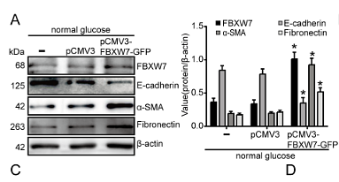
FBXW7 mediates high glucose-induced epithelial to mesenchymal transition via KLF5 in renal tubular cells of diabetic kidney disease
Author: Juan Li, Keqi Jia, Wenjie Wang, Yingxue Pang, Hui Wang, Jun Hao, Dong Zhao, Fan Li
PMID: 40010183

Journal: Tissue & Cell
Application: WB
Reactivity: Human
Publish date: 2025 Feb
-
Citation
-
Silencing CXCR6 promotes epithelial-mesenchymal transition and invasion in colorectal cancer by activating the VEGFA/PI3K/AKT/mTOR pathway
Author: Zhuo Liu,et al
PMID: 39500082
Journal: International Immunopharmacology
Application: IHC-P
Reactivity: Mouse
Publish date: 2024 Nov
-
Citation
-
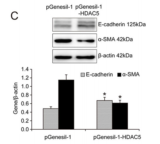
METTL14-regulated PI3K/Akt signaling pathway via PTEN affects HDAC5-mediated epithelial–mesenchymal transition of renal tubular cells in diabetic kidney disease
Author: Xu, Z., Jia, K., Wang, H., Gao, F., Zhao, S., Li, F., & Hao, J.
PMID: 33414476

Journal: Cell Death & Disease
Application: WB
Reactivity: Mouse
Publish date: 2021 Jan
-
Citation
Alternative Products
Products with the same target and pathway
E-Cadherin Recombinant Rabbit Monoclonal Antibody [PSH13-75] - BSA and Azide free
Application: WB,IHC-Fr,IHC-P,IF-Cell,FC,IP
Reactivity: Human,Mouse,Rat
Conjugate: unconjugated
E-Cadherin Mouse Monoclonal Antibody [A0-G11-2]
Application: WB,IHC-P,IF-Tissue
Reactivity: Human,Mouse,Rat
Conjugate: unconjugated
iFluor™ 488 Conjugated E-Cadherin Recombinant Rabbit Monoclonal Antibody [SY0287]
Application: IF-Cell,FC
Reactivity: Human
Conjugate: iFluor™ 488
E-Cadherin Rabbit Polyclonal Antibody
Application: WB,IF-Cell,FC
Reactivity: Human,Mouse
Conjugate: unconjugated
E-Cadherin Recombinant Rabbit Monoclonal Antibody [SY0287] - BSA and Azide free
Application: WB,IF-Cell,IF-Tissue,IHC-P,FC,IP
Reactivity: Human,Cynomolgus monkey
Conjugate: unconjugated
iFluor™ 594 Conjugated E-Cadherin Recombinant Rabbit Monoclonal Antibody [SY0287]
Application: IF-Tissue
Reactivity: Human
Conjugate: iFluor™ 594
E-Cadherin Recombinant Mouse Monoclonal Antibody
Application: mIHC
Reactivity: Human
Conjugate: unconjugated
E-Cadherin Recombinant Rabbit Monoclonal Antibody [PSH13-75]
Application: WB,IHC-Fr,IHC-P,IF-Cell,FC,IP
Reactivity: Human,Mouse,Rat
Conjugate: unconjugated
E-Cadherin Recombinant Rabbit Monoclonal Antibody [SY0287]
Application: WB,IF-Cell,IF-Tissue,IHC-P,FC,IP,mIHC
Reactivity: Human,Cynomolgus monkey
Conjugate: unconjugated
E-Cadherin Recombinant Mouse Monoclonal Antibody [A0-G11-2-R] - BSA and Azide free
Application: WB,IHC-P
Reactivity: Human,Mouse,Rat
Conjugate: unconjugated
E-Cadherin Recombinant Mouse Monoclonal Antibody [A0-G11-2-R]
Application: WB,IHC-P,mIHC
Reactivity: Human,Mouse,Rat
Conjugate: unconjugated
E-Cadherin Mouse Monoclonal Antibody [3-F9]
Application: IHC-P
Reactivity: Human,Mouse
Conjugate: unconjugated
E-Cadherin Recombinant Rabbit Monoclonal Antibody
Application: mIHC
Reactivity: Human
Conjugate: unconjugated


