Cytokeratin 8 Rabbit Polyclonal Antibody
Overview
Product Name
Cytokeratin 8 Rabbit Polyclonal Antibody
Antibody Type
Rabbit Polyclonal Antibody
Immunogen
Recombinant protein within Human Cytokeratin 8 aa 380-483.
Species Reactivity
Human
Validated Applications
WB, IF-Cell, IHC-P, FC
Molecular Weight
53/56 kDa
Positive Control
K562, LOVO, human liver tissue, human breast cancer tissue, human kidney tissue, MCF-7.
Conjugation
unconjugated
RRID
Product Features
Form
Liquid
Concentration
1ug/ul
Storage Instructions
Store at +4℃ after thawing. Aliquot store at -20℃. Avoid repeated freeze / thaw cycles.
Storage Buffer
1*PBS (pH7.4), 0.2% BSA, 50% Glycerol. Preservative: 0.05% Sodium Azide.
Isotype
IgG
Purification Method
Immunogen affinity purified.
Application Dilution
-
WB
-
1:500-1:2,000
-
IF-Cell
-
1:50
-
IHC-P
-
1:50-1:200
-
FC
-
1:50-1:100
Target
Function
Cytokeratins comprise a diverse group of intermediate filament proteins (IFPs) that are expressed as pairs in both keratinized and non-keratinized epithelial tissue. Cytokeratins play a critical role in differentiation and tissue specialization and function to maintain the overall structural integrity of epithelial cells. They have been found to be useful markers of tissue differentiation, which is directly applicable to the characterization of malignant tumors. Cytokeratin 8 expression is seen in epithelium and epithelium-derived tumors. The Cytokeratin 8 and 18 pair are normally expressed in simple epithelia, but not in stratified epithelial cells. Research indicates that squamous cell carcinomas derived from stratified epithelia show abnormal expression of Cytokeratin 8 and 18, although it is not known whether these proteins contribute to the malignant phenotype of the cells. Expression of Cytokeratin 8 and 18 in oral squamous cell carcinomas is an independent prognostic marker that indicates a poor prognosis. Cytokeratin 8 expression correlates with malignancy in leukoplakia and carcinomas of the head and neck; it is expressed in all non-small-cell lung cancers. Cytokeratin 8 has been shown to possess extracellular epitopes on tumor cells, which may represent valuable targets for therapy.
Background References
1. Stone M R et al. Specific interaction of the actin-binding domain of dystrophin with intermediate filaments containing keratin 19. Mol Biol Cell 16:4280-4293 (2005).
2. Barrett A W et al. An immunohistological study of cytokeratin 20 in human and mammalian oral epithelium. Arch Oral Biol 45:879-887 (2000).
Sequence Similarity
Belongs to the intermediate filament family.
Tissue Specificity
Observed in muscle fibers accumulating in the costameres of myoplasm at the sarcolemma membrane in structures that contain dystrophin and spectrin. Expressed in gingival mucosa and hard palate of the oral cavity.
Post-translational Modification
Phosphorylation on serine residues is enhanced during EGF stimulation and mitosis. Ser-74 phosphorylation plays an important role in keratin filament reorganization.; O-glycosylated. O-GlcNAcylation at multiple sites increases solubility, and decreases stability by inducing proteasomal degradation.; O-glycosylated (O-GlcNAcylated), in a cell cycle-dependent manner.
Subcellular Location
Cytoplasm. Nucleus.
UNIPROT
Synonyms
CARD2 antibody
CK 8 antibody
CK-8 antibody
CK8 antibody
CYK8 antibody
CYKER antibody
Cytokeratin endo A antibody
Cytokeratin-8 antibody
DreK8 antibody
EndoA antibody
ExpandCARD2 antibody
CK 8 antibody
CK-8 antibody
CK8 antibody
CYK8 antibody
CYKER antibody
Cytokeratin endo A antibody
Cytokeratin-8 antibody
DreK8 antibody
EndoA antibody
K2C8 antibody
K2C8_HUMAN antibody
K8 antibody
Keratin 8 antibody
Keratin type II cytoskeletal 8 antibody
Keratin, type II cytoskeletal 8 antibody
Keratin-8 antibody
KO antibody
Krt 2.8 antibody
KRT8 antibody
MGC118110 antibody
MGC174782 antibody
MGC53564 antibody
MGC85764 antibody
sb:cb186 antibody
Type-II keratin Kb8 antibody
CollapseImages
-
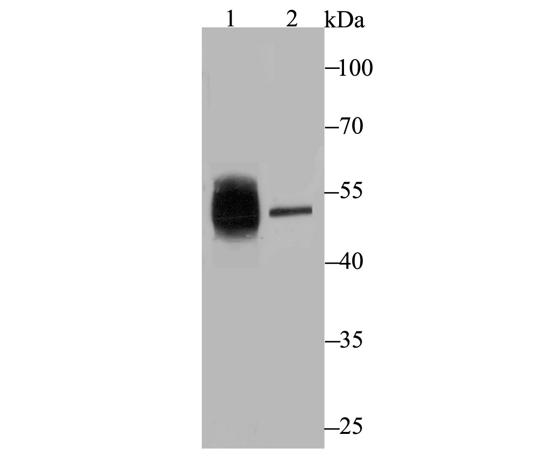
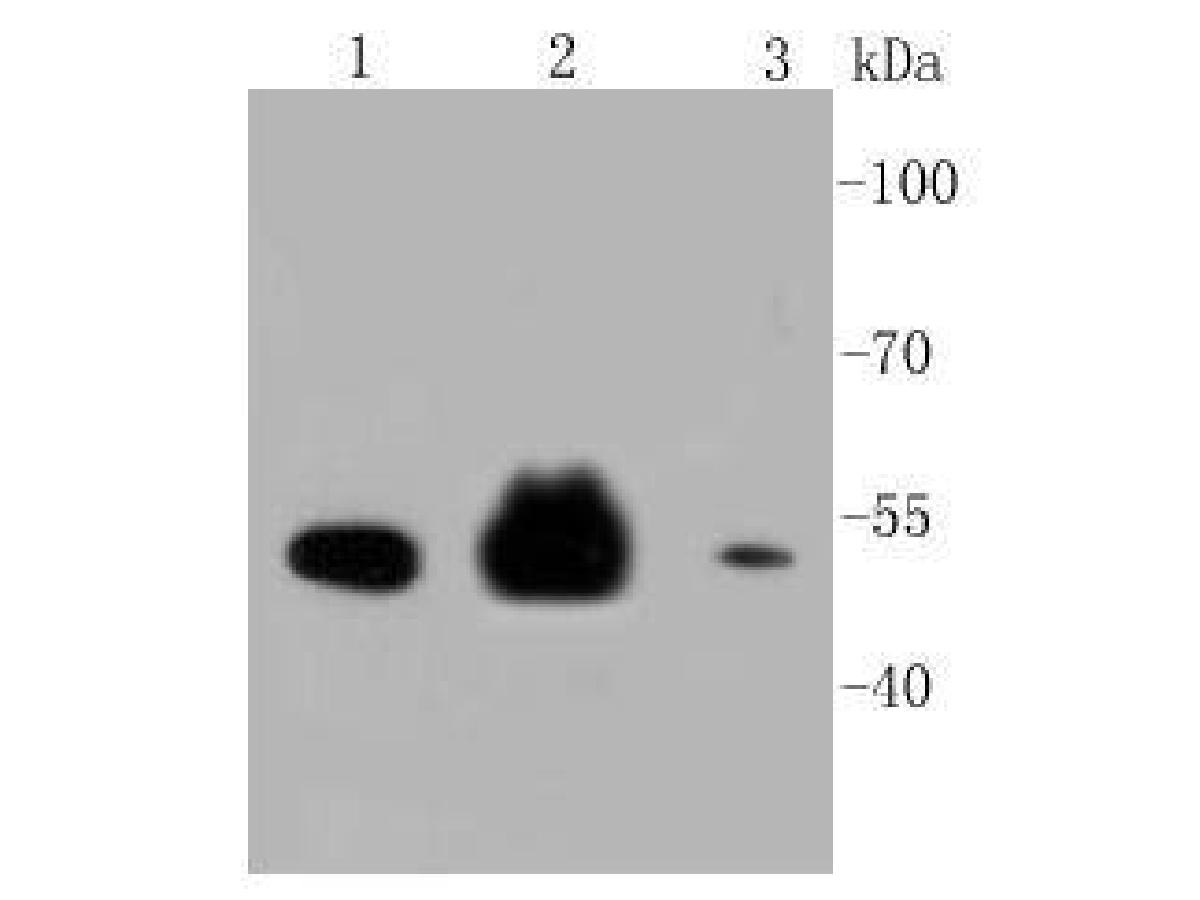
Western blot analysis of Cytokeratin 8 on K562 lysate. Proteins were transferred to a PVDF membrane and blocked with 5% BSA in PBS for 1 hour at room temperature. The primary antibody was used at a 1:2,000 dilution in 5% BSA at room temperature for 2 hours. Goat Anti-Rabbit IgG - HRP Secondary Antibody (HA1001) at 1:5,000 dilution was used for 1 hour at room temperature.
-
ICC staining Cytokeratin 8 in LOVO cells (green). Formalin fixed cells were permeabilized with 0.1% Triton X-100 in TBS for 10 minutes at room temperature and blocked with 1% Blocker BSA for 15 minutes at room temperature. Cells were probed with Cytokeratin 8 at a dilution of 1:50 for 1 hour at room temperature, washed with PBS. Alexa Fluorc™ 488 Goat anti-Rabbit IgG was used as the secondary antibody at 1/100 dilution. The nuclear counter stain is DAPI (blue).
-
Immunohistochemical analysis of paraffin-embedded human liver tissue using anti-Cytokeratin 8 antibody. The section was pre-treated using heat mediated antigen retrieval with Tris-EDTA buffer (pH 8.0-8.4) for 20 minutes.The tissues were blocked in 5% BSA for 30 minutes at room temperature, washed with ddH2O and PBS, and then probed with the antibody (ER1803-87) at 1/50 dilution, for 30 minutes at room temperature and detected using an HRP conjugated compact polymer system. DAB was used as the chromogen. Counter stained with hematoxylin and mounted with DPX.
-
Immunohistochemical analysis of paraffin-embedded human breast cancer tissue using anti-Cytokeratin 8 antibody. The section was pre-treated using heat mediated antigen retrieval with Tris-EDTA buffer (pH 8.0-8.4) for 20 minutes.The tissues were blocked in 5% BSA for 30 minutes at room temperature, washed with ddH2O and PBS, and then probed with the antibody (ER1803-87) at 1/200 dilution, for 30 minutes at room temperature and detected using an HRP conjugated compact polymer system. DAB was used as the chromogen. Counter stained with hematoxylin and mounted with DPX.
-
Immunohistochemical analysis of paraffin-embedded human kidney tissue using anti-Cytokeratin 8 antibody. The section was pre-treated using heat mediated antigen retrieval with Tris-EDTA buffer (pH 8.0-8.4) for 20 minutes.The tissues were blocked in 5% BSA for 30 minutes at room temperature, washed with ddH2O and PBS, and then probed with the antibody (ER1803-87) at 1/200 dilution, for 30 minutes at room temperature and detected using an HRP conjugated compact polymer system. DAB was used as the chromogen. Counter stained with hematoxylin and mounted with DPX.
-
Flow cytometric analysis of Cytokeratin 8 was done on MCF-7 cells. The cells were fixed, permeabilized and stained with Cytokeratin 8 antibody at 1/100 dilution (red) compared with an unlabelled control (cells without incubation with primary antibody; black). After incubation of the primary antibody on room temperature for an hour, the cells was stained with a Alexa Fluor™ 488-conjugated goat anti-Rabbit IgG Secondary antibody at 1/500 dilution for 30 minutes.
Please note: All products are "FOR RESEARCH USE ONLY AND ARE NOT INTENDED FOR DIAGNOSTIC OR THERAPEUTIC USE"
Alternative Products
Cytokeratin 8 Recombinant Rabbit Monoclonal Antibody [SU0338]
Application: WB,IF-Cell,IF-Tissue,IHC-P,IP,FC
Reactivity: Human,Mouse
Conjugate: unconjugated
Products with the same target and pathway
iFluor™ 647 Conjugated Cytokeratin 8 Recombinant Rabbit Monoclonal Antibody [SU0338]
Application: IF-Cell,IF-Tissue
Reactivity: Human,Mouse
Conjugate: iFluor™ 647
Cytokeratin 8 Rabbit Polyclonal Antibody
Application: WB,IF-Cell,IHC-P,FC
Reactivity: Human,Mouse,Rat
Conjugate: unconjugated
Cytokeratin 8
Application:
Reactivity:
Conjugate:
Cytokeratin 8 Rabbit Polyclonal Antibody
Application: WB,IF-Cell,IHC-P,FC
Reactivity: Human,Mouse,Rat
Conjugate: unconjugated
Cytokeratin 8 Recombinant Rabbit Monoclonal Antibody [SU0338]
Application: WB,IF-Cell,IF-Tissue,IHC-P,IP,FC
Reactivity: Human,Mouse
Conjugate: unconjugated
iFluor™ 488 Conjugated Cytokeratin 8 Recombinant Rabbit Monoclonal Antibody [SU0338]
Application: IF-Tissue,IF-Cell
Reactivity: Human
Conjugate: iFluor™ 488
Cytokeratin 8 Mouse Monoclonal Antibody [A1-B11]
Application: WB,IF-Cell,IHC-P,FC
Reactivity: Human,Mouse,Rat
Conjugate: unconjugated
iFluor™ 594 Conjugated Cytokeratin 8 Recombinant Rabbit Monoclonal Antibody [SU0338]
Application: IF-Cell,IF-Tissue,FC
Reactivity: Human,Mouse
Conjugate: iFluor™ 594