CD38 Recombinant Rabbit Monoclonal Antibody [PD01-49]
Specification
Catalog# HA721268
CD38 Recombinant Rabbit Monoclonal Antibody [PD01-49]
-
WB
-
IHC-P
-
IF-Cell
-
FC
-
mIHC
-
IF-Tissue
-
Human
-
unconjugated
Overview
Product Name
CD38 Recombinant Rabbit Monoclonal Antibody [PD01-49]
Antibody Type
Recombinant Rabbit monoclonal Antibody
Immunogen
Synthetic peptide within human CD38 aa 250-300.
Species Reactivity
Human
Validated Applications
WB, IHC-P, IF-Cell, FC, mIHC, IF-Tissue
Molecular Weight
Predicted band size: 34 kDa
Positive Control
Daudi cell lysate, Ramos cell lysate, A549, human tonsil tissue, THP-1, human appendix tissue, human colon cancer tissue, human prostate tissue, human thymus tissue.
Conjugation
unconjugated
Clone Number
PD01-49
RRID
Product Features
Form
Liquid
Storage Instructions
Shipped at 4℃. Store at +4℃ short term (1-2 weeks). It is recommended to aliquot into single-use upon delivery. Store at -20℃ long term.
Storage Buffer
PBS (pH7.4), 0.1% BSA, 40% Glycerol. Preservative: 0.05% Sodium Azide.
Isotype
IgG
Purification Method
Protein A affinity purified.
Application Dilution
-
WB
-
1:2,000
-
IHC-P
-
1:1,000
-
IF-Cell
-
1:50
-
FC
-
1:1,000
-
mIHC
-
1:1,000-1:4,000
-
IF-Tissue
-
1:500
Applications in Publications
Species in Publications
| Human | See 1 publications below |
Target
Function
CD38 is a type II integral membrane glycoprotein which is present on early B and T cell lineages and activated B and T cells but is absent from most mature resting peripheral lymphocytes. CD38 is also found on thymocytes, pre-B cells, germinal center B cells, mitogen-activated T cells, monocytes and Ig-secreting plasma cells. CD38 acts as a NAD glycohydrolase in T lymphocytes. On hematopoietic cells CD38 induces activation, proliferation, and differentiation of mature T and B cells and mediates apoptosis of myeloid and lymphoid progenitor cells. In addition to acting as a signaling receptor, CD38 is also an enzyme capable of producing several calcium-mobilizing metabolites, including cyclic adenosine diphosphate ribose (cADPR). CD38 also plays a role in maintaining survival of an invariant NK T (iNKT) cell subset that preferentially contributes to the maintenance of immunological tolerance.
Background References
1. Yang Q et al. NADase CD38 is a key determinant of ovarian aging. Nat Aging. 2024 Jan
2. Yu S et al. CD38-Targeting Peptide Vaccine Ameliorates Aging-Associated Phenotypes in Mice. Aging Cell. 2025 Sep
Subcellular Location
Membrane.
UNIPROT
Synonyms
Acute lymphoblastic leukemia cells antigen CD38 antibody
ADP ribosyl cyclase 1 antibody
ADP ribosyl cyclase antibody
ADP ribosyl cyclase/cyclic ADP-ribose hydrolase antibody
ADP-ribosyl cyclase 1 antibody
ADPRC 1 antibody
ADPRC1 antibody
cADPr hydrolase 1 antibody
CD 38 antibody
CD38 antibody
ExpandAcute lymphoblastic leukemia cells antigen CD38 antibody
ADP ribosyl cyclase 1 antibody
ADP ribosyl cyclase antibody
ADP ribosyl cyclase/cyclic ADP-ribose hydrolase antibody
ADP-ribosyl cyclase 1 antibody
ADPRC 1 antibody
ADPRC1 antibody
cADPr hydrolase 1 antibody
CD 38 antibody
CD38 antibody
CD38 antigen (p45) antibody
CD38 antigen antibody
CD38 molecule antibody
Cd38-rs1 antibody
CD38_HUMAN antibody
CD38H antibody
Cyclic ADP ribose hydrolase antibody
Cyclic ADP ribose hydrolase 1 antibody
Cyclic ADP-ribose hydrolase 1 antibody
EC 3.2.2.5 antibody
Ecto nicotinamide adenine dinucleotide glycohydrolase antibody
I-19 antibody
I19 (mouse) antibody
Lymphocyte differentiation antigen CD38 antibody
NAD(+) nucleosidase antibody
NIM-R5 antigen antibody
NIMR5 antigen (mouse) antibody
OTTHUMP00000158633 antibody
OTTHUMP00000217743 antibody
p45 antibody
T10 antibody
CollapseImages
-

☑ Relative expression (RE)
Western blot analysis of CD38 on different lysates with Rabbit anti-CD38 antibody (HA721268) at 1/2,000 dilution.
Lane 1: Daudi cell lysate
Lane 2: Ramos cell lysate
Lane 3: HepG2 cell lysate (negative)
Lysates/proteins at 20 µg/Lane.
Predicted band size: 34 kDa
Observed band size: 45 kDa
Exposure time: 21 seconds; ECL: K1801;
4-20% SDS-PAGE gel.
Proteins were transferred to a PVDF membrane and blocked with 5% NFDM/TBST for 1 hour at room temperature. The primary antibody (HA721268) at 1/2,000 dilution was used in primary antibody dilution (K1803) at 4℃ overnight. Goat Anti-Rabbit IgG - HRP Secondary Antibody (HA1001) at 1/50,000 dilution was used for 1 hour at room temperature. -

Fluorescence multiplex immunohistochemical analysis of Human tonsil (Formalin/PFA-fixed paraffin-embedded sections). Panel A: the merged image of anti-CD68 (HA601115, Red), anti-CD38 (HA721268, Green), anti-CD23 (HA721139, White), anti-CD11C (ET1606-19, Cyan), anti-CD45 (ET7111-03, Magenta) and anti-CD20 (HA721138, Yellow) on tonsil. Panel B: anti-CD68 stained on Macrophage. Panel C: anti-CD38 stained on lymphocyte subsets. Panel D: anti-CD11C stained on dendritic cells. Panel E: CD45 stained on lymphocytes. Panel F: anti-CD20 stained on B cells. Panel G: anti-CD23 stained on follicular dendritic cells. HRP Conjugated UltraPolymer Goat Polyclonal Antibody HA1119/HA1120 was used as a secondary antibody. The immunostaining was performed with the Sequential Immuno-staining Kit (IRISKit™MH010101, www.luminiris.cn). The section was incubated in six rounds of staining: in the order of HA601115 (1/2,000 dilution), HA721268 (1/1,000 dilution), ET1606-19 (1/1,000 dilution), ET7111-03 (1/500 dilution), HA721138 (1/2,000 dilution) and HA721139 (1/800 dilution) for 20 mins at room temperature. Each round was followed by a separate fluorescent tyramide signal amplification system. Heat mediated antigen retrieval with Tris-EDTA buffer (pH 9.0) for 30 mins at 95℃. DAPI (blue) was used as a nuclear counter stain. Image acquisition was performed with Olympus VS200 Slide Scanner.
-

Fluorescence multiplex immunohistochemical analysis of human tonsil (Formalin/PFA-fixed paraffin-embedded sections). Panel A: the merged image of anti-CD20 (HA721138, Cyan), anti-CD38 (HA721268, Violet) and anti-CD57 (HA601114, Yellow) on tonsil. HRP Conjugated UltraPolymer Goat Polyclonal Antibody HA1119/HA1120 was used as a secondary antibody. The immunostaining was performed with the Sequential Immuno-staining Kit (IRISKit™MH010101, www.luminiris.cn). The section was incubated in three rounds of staining: in the order of HA721138 (1/2,000 dilution), HA721268 (1/1,000 dilution) and HA601114 (1/1,000 dilution) for 20 mins at room temperature. Each round was followed by a separate fluorescent tyramide signal amplification system. Heat mediated antigen retrieval with Tris-EDTA buffer (pH 9.0) for 30 mins at 95℃. DAPI (blue) was used as a nuclear counter stain. Image acquisition was performed with Zeiss Observer 7 Inverted Fluorescence Microscope.
-

Immunocytochemistry analysis of A549 cells labeling CD38 with Rabbit anti-CD38 antibody (HA721268) at 1/50 dilution.
Cells were fixed in 4% paraformaldehyde for 10 minutes at 37 ℃, permeabilized with 0.05% Triton X-100 in PBS for 20 minutes, and then blocked with 2% negative goat serum for 30 minutes at room temperature. Cells were then incubated with Rabbit anti-CD38 antibody (HA721268) at 1/50 dilution in 2% negative goat serum overnight at 4 ℃. Goat Anti-Rabbit IgG H&L (iFluor™ 488, HA1121) was used as the secondary antibody at 1/1,000 dilution. PBS instead of the primary antibody was used as the secondary antibody only control. Nuclear DNA was labelled in blue with DAPI. -

Immunohistochemical analysis of paraffin-embedded human tonsil tissue with Rabbit anti-CD38 antibody (HA721268) at 1/2,000 dilution.
The section was pre-treated using heat mediated antigen retrieval with Tris-EDTA buffer (pH 9.0) for 20 minutes. The tissues were blocked in 1% BSA for 20 minutes at room temperature, washed with ddH2O and PBS, and then probed with the primary antibody (HA721268) at 1/2,000 dilution for 1 hour at room temperature. The detection was performed using an HRP conjugated compact polymer system. DAB was used as the chromogen. Tissues were counterstained with hematoxylin and mounted with DPX. -

Flow cytometric analysis of THP-1 cells labeling CD38.
Cells were washed twice with cold PBS and resuspend. Then stained with the primary antibody (HA721268, 1μg/mL) (red) compared with Rabbit IgG Isotype Control (green). After incubation of the primary antibody at +4℃ for an hour, the cells were stained with a iFluor™ 488 conjugate-Goat anti-Rabbit IgG Secondary antibody (HA1121) at 1/1,000 dilution for 30 minutes at +4℃. Unlabelled sample was used as a control (cells without incubation with primary antibody; black). -

Immunofluorescence analysis of paraffin-embedded human appendix tissue labeling CD38 with Rabbit anti-CD38 antibody (HA721268) at 1/500 dilution.
The section was pre-treated using heat mediated antigen retrieval with Tris-EDTA buffer (pH 9.0) for 20 minutes. The tissues were blocked in 10% negative goat serum for 1 hour at room temperature, washed with PBS, and then probed with the primary antibody (HA721268, green) at 1/500 dilution overnight at 4 ℃, washed with PBS. Goat Anti-Rabbit IgG H&L (iFluor™ 488, HA1121) was used as the secondary antibody at 1/1,000 dilution. Nuclei were counterstained with DAPI (blue). -

Immunohistochemical analysis of paraffin-embedded human appendix tissue with Rabbit anti-CD38 antibody (HA721268) at 1/2,000 dilution.
The section was pre-treated using heat mediated antigen retrieval with Tris-EDTA buffer (pH 9.0) for 20 minutes. The tissues were blocked in 1% BSA for 20 minutes at room temperature, washed with ddH2O and PBS, and then probed with the primary antibody (HA721268) at 1/2,000 dilution for 1 hour at room temperature. The detection was performed using an HRP conjugated compact polymer system. DAB was used as the chromogen. Tissues were counterstained with hematoxylin and mounted with DPX. -

Immunohistochemical analysis of paraffin-embedded human colon cancer tissue with Rabbit anti-CD38 antibody (HA721268) at 1/2,000 dilution.
The section was pre-treated using heat mediated antigen retrieval with Tris-EDTA buffer (pH 9.0) for 20 minutes. The tissues were blocked in 1% BSA for 20 minutes at room temperature, washed with ddH2O and PBS, and then probed with the primary antibody (HA721268) at 1/2,000 dilution for 1 hour at room temperature. The detection was performed using an HRP conjugated compact polymer system. DAB was used as the chromogen. Tissues were counterstained with hematoxylin and mounted with DPX. -

Immunohistochemical analysis of paraffin-embedded human prostate tissue with Rabbit anti-CD38 antibody (HA721268) at 1/2,000 dilution.
The section was pre-treated using heat mediated antigen retrieval with Tris-EDTA buffer (pH 9.0) for 20 minutes. The tissues were blocked in 1% BSA for 20 minutes at room temperature, washed with ddH2O and PBS, and then probed with the primary antibody (HA721268) at 1/2,000 dilution for 1 hour at room temperature. The detection was performed using an HRP conjugated compact polymer system. DAB was used as the chromogen. Tissues were counterstained with hematoxylin and mounted with DPX. -

Immunohistochemical analysis of paraffin-embedded human thymus tissue with Rabbit anti-CD38 antibody (HA721268) at 1/2,000 dilution.
The section was pre-treated using heat mediated antigen retrieval with Tris-EDTA buffer (pH 9.0) for 20 minutes. The tissues were blocked in 1% BSA for 20 minutes at room temperature, washed with ddH2O and PBS, and then probed with the primary antibody (HA721268) at 1/2,000 dilution for 1 hour at room temperature. The detection was performed using an HRP conjugated compact polymer system. DAB was used as the chromogen. Tissues were counterstained with hematoxylin and mounted with DPX. -

☑ Relative expression (RE)
Immunohistochemical analysis of paraffin-embedded human liver tissue (negative) with Rabbit anti-CD38 antibody (HA721268) at 1/2,000 dilution.
The section was pre-treated using heat mediated antigen retrieval with Tris-EDTA buffer (pH 9.0) for 20 minutes. The tissues were blocked in 1% BSA for 20 minutes at room temperature, washed with ddH2O and PBS, and then probed with the primary antibody (HA721268) at 1/2,000 dilution for 1 hour at room temperature. The detection was performed using an HRP conjugated compact polymer system. DAB was used as the chromogen. Tissues were counterstained with hematoxylin and mounted with DPX. -

Immunohistochemical analysis of paraffin-embedded human tonsil tissue with Rabbit anti-CD38 antibody (HA721268) at 1/1,000 dilution.
Heat mediated antigen retrieval with Tris-EDTA buffer (pH 9.0, epitope retrieval solution 2) for 20 mins. The section was incubated with HA721268 for 30 mins at room temperature. The immunostaining was performed on a Leica Biosystems BOND® RX instrument. DAB was used as the chromogen. Tissues were counterstained with hematoxylin and mounted with DPX.
Please note: All products are "FOR RESEARCH USE ONLY AND ARE NOT INTENDED FOR DIAGNOSTIC OR THERAPEUTIC USE"
Citation
-
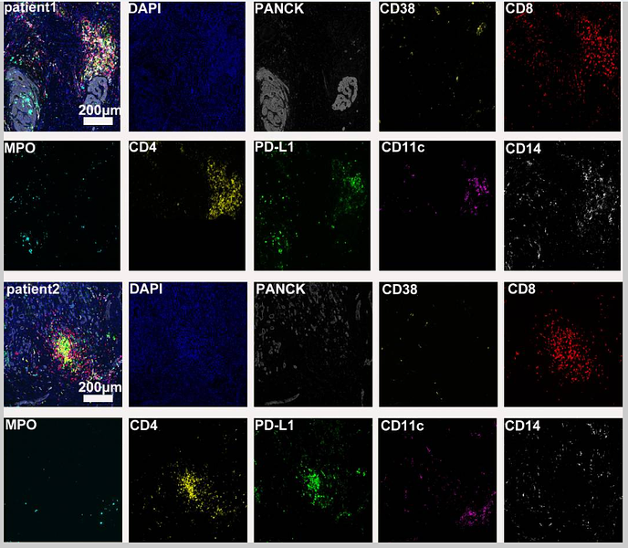
Case report: Diverse immune responses in advanced pancreatic ductal adenocarcinoma treated with immune checkpoint inhibitor-based conversion therapies
Author: Li Xiaoying, Xiao Chaoxin, Li Ruizhen, Zhang Pei, Cao Dan
PMID: 38415262

Journal: Frontiers In Immunology
Application: mIHC
Reactivity: Human
Publish date: 2024 Feb
-
Citation
Products with the same target and pathway
CD38 Recombinant Antibody - Mouse IgG1 (Chimeric)
Application: mIHC
Reactivity: Human
Conjugate: unconjugated
CD38 Recombinant Antibody [PD01-49] - Mouse IgG1 (Chimeric) - BSA and Azide free
Application: WB,IHC-P,FC
Reactivity: Human
Conjugate: unconjugated
CD38 Recombinant Antibody - Rat IgG1 (Chimeric)
Application: mIHC
Reactivity: Human
Conjugate: unconjugated
CD38 Recombinant Rabbit Monoclonal Antibody [JE29-35]
Application: WB,IHC-P,FC,IF-Tissue
Reactivity: Human
Conjugate: unconjugated
CD38 Recombinant Antibody [PD01-49] - Rat IgG1 (Chimeric)
Application: mIHC
Reactivity: Human
Conjugate: unconjugated
CD38 Recombinant Antibody [PD01-49] - Rat IgG1 (Chimeric) - BSA and Azide free
Application:
Reactivity: Human
Conjugate: unconjugated
CD38
Application:
Reactivity:
Conjugate:
CD38 Recombinant Antibody [PD01-49] - Mouse IgG1 (Chimeric)
Application: WB,IHC-P,FC,mIHC
Reactivity: Human
Conjugate: unconjugated
CD38 Recombinant Rabbit Monoclonal Antibody
Application: mIHC
Reactivity: Human
Conjugate: unconjugated
CD38 Recombinant Rabbit Monoclonal Antibody [SN07-20]
Application: WB,IHC-P,FC,IF-Tissue
Reactivity: Human
Conjugate: unconjugated


