Phospho-FAK (Y397) Recombinant Rabbit Monoclonal Antibody [SC54-07]

Specification
Catalog# ET1610-34
Phospho-FAK (Y397) Recombinant Rabbit Monoclonal Antibody [SC54-07]
-
WB
-
IF-Cell
-
IF-Tissue
-
IHC-P
-
Human
-
Mouse
-
Rat
Overview
Product Name
Phospho-FAK (Y397) Recombinant Rabbit Monoclonal Antibody [SC54-07]
Antibody Type
Recombinant Rabbit monoclonal Antibody
Immunogen
Synthetic phospho-peptide corresponding to residues surrounding Tyr397 of human FAK.
Species Reactivity
Human, Mouse, Rat
Validated Applications
WB, IF-Cell, IF-Tissue, IHC-P
Molecular Weight
Predicted band size: 119 kDa
Positive Control
Hela cell lysate, PC-3 cell lysate, NIH/3T3, C6, Hela, mouse spleen tissue, rat spleen tissue.
Conjugation
unconjugated
Clone Number
SC54-07
RRID
Product Features
Form
Liquid
Storage Instructions
Shipped at 4℃. Store at +4℃ short term (1-2 weeks). It is recommended to aliquot into single-use upon delivery. Store at -20℃ long term.
Storage Buffer
1*TBS (pH7.4), 0.05% BSA, 40% Glycerol. Preservative: 0.05% Sodium Azide.
Isotype
IgG
Purification Method
Protein A affinity purified.
Application Dilution
-
WB
-
1:500-1:1,000
-
IF-Cell
-
1:50-1:100
-
IF-Tissue
-
1:50-1:100
-
IHC-P
-
1:200
Applications in Publications
Species in Publications
Target
Function
Activation of integrins in the extracellular matrix (ECM) of eukaryotic cells promotes the formation of membrane adhesion complexes, known as focal adhesions, which can include cytoskeletal proteins and protein tyrosine kinases, such as focal adhesion kinase (FAK). Phosphorylation events occurring within focal adhesions influence numerous processes that include mitogenic signaling, cell survival, and cell motility. FAK is a non-receptor tyrosine kinase that is ubiquitously expressed and highly conserved between species. FAK is recruited by Integrin clusters and variably phosphorylated depending on the effector molecules present in the focal adhesion. Phosphorylation of FAK Tyr 397 decreases during serum starvation, contact inhibition, and cell cycle arrest, all conditions under which activating FAK Tyr 407 phosphorylation increases.
Background References
1. Kuo SW et al. Regulation of the fate of human mesenchymal stem cells by mechanical and stereo-topographical cues provided by silicon nanowires. Biomaterials 33:5013-22 (2012).
2. Lu H et al. IGFBP2/FAK pathway is causally associated with dasatinib resistance in non-small cell lung cancer cells. Mol Cancer Ther 12:2864-73 (2013).
Sequence Similarity
Belongs to the protein kinase superfamily. Tyr protein kinase family. FAK subfamily.
Tissue Specificity
Detected in B and T-lymphocytes. Isoform 1 and isoform 6 are detected in lung fibroblasts (at protein level). Ubiquitous. Expressed in epithelial cells (at protein level).
Post-translational Modification
Phosphorylated on tyrosine residues upon activation, e.g. upon integrin signaling. Tyr-397 is the major autophosphorylation site, but other kinases can also phosphorylate this residue. Phosphorylation at Tyr-397 promotes interaction with SRC and SRC family members, leading to phosphorylation at Tyr-576, Tyr-577 and at additional tyrosine residues. FGR promotes phosphorylation at Tyr-397 and Tyr-576. FER promotes phosphorylation at Tyr-577, Tyr-861 and Tyr-925, even when cells are not adherent. Tyr-397, Tyr-576 and Ser-722 are phosphorylated only when cells are adherent. Phosphorylation at Tyr-397 is important for interaction with BMX, PIK3R1 and SHC1. Phosphorylation at Tyr-925 is important for interaction with GRB2. Dephosphorylated by PTPN11; PTPN11 is recruited to PTK2 via EPHA2 (tyrosine phosphorylated). Microtubule-induced dephosphorylation at Tyr-397 is crucial for the induction of focal adhesion disassembly; this dephosphorylation could be catalyzed by PTPN11 and regulated by ZFYVE21. Phosphorylation on tyrosine residues is enhanced by NTN1 (By similarity).; Sumoylated; this enhances autophosphorylation.
Subcellular Location
Cytoplasm, Nucleus, Cell membrane, Cell junction.
Synonyms
FADK 1 antibody
FADK antibody
FAK related non kinase polypeptide antibody
FAK1 antibody
FAK1_HUMAN antibody
Focal adhesion kinase 1 antibody
Focal adhesion Kinase antibody
Focal adhesion kinase isoform FAK Del33 antibody
Focal adhesion kinase related nonkinase antibody
FRNK antibody
ExpandFADK 1 antibody
FADK antibody
FAK related non kinase polypeptide antibody
FAK1 antibody
FAK1_HUMAN antibody
Focal adhesion kinase 1 antibody
Focal adhesion Kinase antibody
Focal adhesion kinase isoform FAK Del33 antibody
Focal adhesion kinase related nonkinase antibody
FRNK antibody
p125FAK antibody
pp125FAK antibody
PPP1R71 antibody
Protein phosphatase 1 regulatory subunit 71 antibody
Protein tyrosine kinase 2 antibody
Protein-tyrosine kinase 2 antibody
Ptk2 antibody
PTK2 protein tyrosine kinase 2 antibody
CollapseImages
-

Western blot analysis of Phospho-FAK (Y397) on different lysates with Rabbit anti-Phospho-FAK (Y397) antibody (ET1610-34) at 1/500 dilution.
Lane 1: Hela cell lysate
Lane 2: PC-3 cell lysate
Lysates/proteins at 10 µg/Lane.
Predicted band size: 119 kDa
Observed band size: 119 kDa
Exposure time: 2 minutes;
6% SDS-PAGE gel.
Proteins were transferred to a PVDF membrane and blocked with 5% NFDM/TBST for 1 hour at room temperature. The primary antibody (ET1610-34) at 1/500 dilution was used in 5% NFDM/TBST at room temperature for 2 hours. Goat Anti-Rabbit IgG - HRP Secondary Antibody (HA1001) at 1:300,000 dilution was used for 1 hour at room temperature. -

☑ Cell treatment (CT)
Western blot analysis of Phospho-FAK (Y397) on Hela cell lysates.
Lane 1: Hela cells, whole cell lysate, 10ug/lane
Lane 2: Hela cells treated with 2.8ug/ul lambda-PP for 30 minutes, whole cell lysates, 10ug/lane
All lanes :
Anti-Phospho-FAK (Y397) antibody (ET1610-34) at 1:500 dilution. Anti-Hsp90 antibody (ET1605-56) at 1:10,000 dilution. Goat Anti-Rabbit IgG H&L (HRP) (HA1001) at 1/200,000 dilution.
Predicted band size: 119 kDa
Observed band size: 119 kDa
Blocking and diluting buffer: 5% BSA.
Exposure time: 30 seconds -

Immunocytochemistry analysis of NIH/3T3 cells labeling Phospho-FAK (Y397) with Rabbit anti-Phospho-FAK (Y397) antibody (ET1610-34) at 1/50 dilution.
Cells were fixed in 4% paraformaldehyde for 15 minutes at room temperature, permeabilized with 0.1% Triton X-100 in PBS for 15 minutes at room temperature, then blocked with 1% BSA in 10% negative goat serum for 1 hour at room temperature. Cells were then incubated with Rabbit anti-Phospho-FAK (Y397) antibody (ET1610-34) at 1/50 dilution in 1% BSA in PBST overnight at 4 ℃. Goat Anti-Rabbit IgG H&L (iFluor™ 488, HA1121) was used as the secondary antibody at 1/1,000 dilution. PBS instead of the primary antibody was used as the secondary antibody only control. Nuclear DNA was labelled in blue with DAPI.
Beta tubulin (HA601187, red) was stained at 1/100 dilution overnight at +4℃. Goat Anti-Mouse IgG H&L (iFluor™ 594, HA1126) was used as the secondary antibody at 1/1,000 dilution. -

Immunocytochemistry analysis of C6 cells labeling Phospho-FAK (Y397) with Rabbit anti-Phospho-FAK (Y397) antibody (ET1610-34) at 1/100 dilution.
Cells were fixed in 4% paraformaldehyde for 15 minutes at room temperature, permeabilized with 0.1% Triton X-100 in PBS for 15 minutes at room temperature, then blocked with 1% BSA in 10% negative goat serum for 1 hour at room temperature. Cells were then incubated with Rabbit anti-Phospho-FAK (Y397) antibody (ET1610-34) at 1/100 dilution in 1% BSA in PBST overnight at 4 ℃. Goat Anti-Rabbit IgG H&L (iFluor™ 488, HA1121) was used as the secondary antibody at 1/1,000 dilution. PBS instead of the primary antibody was used as the secondary antibody only control. Nuclear DNA was labelled in blue with DAPI.
Beta tubulin (HA601187, red) was stained at 1/100 dilution overnight at +4℃. Goat Anti-Mouse IgG H&L (iFluor™ 594, HA1126) was used as the secondary antibody at 1/1,000 dilution. -

ICC staining of Phospho-FAK (Y397) in Hela cells (green). Formalin fixed cells were permeabilized with 0.1% Triton X-100 in TBS for 10 minutes at room temperature and blocked with 1% Blocker BSA for 15 minutes at room temperature. Cells were probed with the primary antibody (ET1610-34, 1/50) for 1 hour at room temperature, washed with PBS. Alexa Fluor®488 Goat anti-Rabbit IgG was used as the secondary antibody at 1/1,000 dilution. The nuclear counter stain is DAPI (blue).
-

Immunohistochemical analysis of paraffin-embedded mouse spleen tissue with Rabbit anti-Phospho-FAK (Y397) antibody (ET1610-34) at 1/200 dilution.
The section was pre-treated using heat mediated antigen retrieval with Tris-EDTA buffer (pH 9.0) for 20 minutes. The tissues were blocked in 1% BSA for 20 minutes at room temperature, washed with ddH2O and PBS, and then probed with the primary antibody (ET1610-34) at 1/200 dilution for 1 hour at room temperature. The detection was performed using an HRP conjugated compact polymer system. DAB was used as the chromogen. Tissues were counterstained with hematoxylin and mounted with DPX. -

Immunohistochemical analysis of paraffin-embedded rat spleen tissue with Rabbit anti-Phospho-FAK (Y397) antibody (ET1610-34) at 1/200 dilution.
The section was pre-treated using heat mediated antigen retrieval with Tris-EDTA buffer (pH 9.0) for 20 minutes. The tissues were blocked in 1% BSA for 20 minutes at room temperature, washed with ddH2O and PBS, and then probed with the primary antibody (ET1610-34) at 1/200 dilution for 1 hour at room temperature. The detection was performed using an HRP conjugated compact polymer system. DAB was used as the chromogen. Tissues were counterstained with hematoxylin and mounted with DPX.
Please note: All products are "FOR RESEARCH USE ONLY AND ARE NOT INTENDED FOR DIAGNOSTIC OR THERAPEUTIC USE"
Citation
-
Local abaloparatide administration promotes in situ alveolar bone augmentation via FAK-mediated periosteal osteogenesis
Author: Yu Li
PMID: 40890141
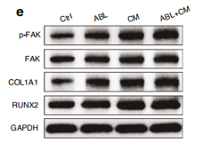
Journal: International Journal Of Oral Science
Application: IHC,WB
Reactivity: Rat
Publish date: 2025 Sept
-
Citation
-
Collagen Glycation-Mediated Mechanical Stress Aggravates Ischemia-Reperfusion injury
Author: Jing Yang, Yixuan Li, Xiaoxiao Fan, Tong Zhang, Junhai Pan, Si Shen, Cheng Zhong, Duguang Li, Yi Zhang, Guoqiao Chen, Wei Ji, Shuqi Wu, Shengxi Jin, Xiaolong Liu, Qiming Xia, Peilin Yu, Wei Yang, Yang Gao, Liangfei Tian, Hui Lin
PMID: 40484296
Journal: Acta Biomaterialia
Application: WB
Reactivity: Human
Publish date: 2025 Jun
-
Citation
-
Integrin β5 interacts with G3BP1 through activating FAK/Src signaling pathway to promote gastric carcinogenesis
Author: Liu Dongliang, Cao Jianjian, Hu Kongwang, Peng Zhiwei
PMID: 40764649
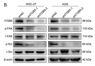
Journal: Scientific Reports
Application: WB
Reactivity: Human
Publish date: 2025 Aug
-
Citation
-
IKIP downregulates THBS1/FAK signaling to suppress migration and invasion by glioblastoma cells
Author: Zhu Zhaoying,et al
PMID: 38948026
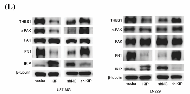
Journal: Oncology Research
Application: WB
Reactivity: Human
Publish date: 2024 Jul
-
Citation
-
The EPRS-ATF4-COLI pathway axis is a potential target for anaplastic thyroid carcinoma therapy
Author: Li Mi, Jiaye Liu, Yujie Zhang, Anping Su, Minghai Tang, Zhichao Xing, Ting He, Tao Wei, Zhihui Li, Wenshuang Wu
PMID: 38704915
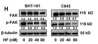
Journal: Phytomedicine
Application: WB
Reactivity: Human
Publish date: 2024 Apr
-
Citation
-
The osteogenic response to chirality-patterned surface potential distribution of CFO/P(VDF-TrFE) membranes
Author: Jiamin Zhang, Xuzhao He, Zhiyuan Zhou, Xiaoyi Chen, Jiaqi Shao, Donghua Huang, Lingqing Dong, Jun Lin, Huiming Wang, Wenjian Weng, Kui Cheng
PMID: 35791864
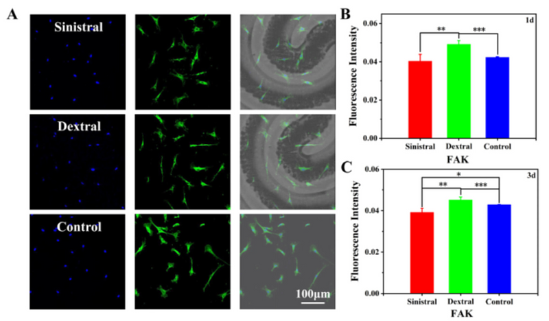
Journal: Biomaterials Science
Application: IF
Reactivity: Rat
Publish date: 2022 Jun
-
Citation
-
Design, synthesis and biological evaluation of novel FAK inhibitors with better selectivity over IR than TAE226
Author:
PMID: 35452915
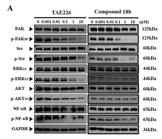
Journal: Bioorganic Chemistry
Application: WB
Reactivity: Human
Publish date: 2022 Jul
-
Citation
-
Cytosolic phospholipase A2α modulates cell-matrix adhesion via the FAK/paxillin pathway in hepatocellular carcinoma
Author: Hua Guo
PMID: 31516757
Journal: Cancer Biology & Medicine
Application: WB,IHC-P,IF
Reactivity: Human
Publish date: 2019 May
-
Citation
Products with the same target and pathway
FAK Rabbit Polyclonal Antibody
Application: WB,IP,ICC,IHC-P
Reactivity: Human,Mouse,Rat
Conjugate: unconjugated
Phospho-FAK (Y397) Recombinant Rabbit Monoclonal Antibody [SC54-07] - BSA and Azide free
Application: WB,IF-Cell,IF-Tissue,IHC-P
Reactivity: Human,Mouse,Rat
Conjugate: unconjugated
FAK Recombinant Rabbit Monoclonal Antibody [SR46-04] - BSA and Azide free
Application: WB,IF-Cell,IF-Tissue,IHC-P,FC,IP
Reactivity: Human,Mouse,Rat
Conjugate: unconjugated
FAK Recombinant Rabbit Monoclonal Antibody [SR46-04]
Application: WB,IF-Cell,IF-Tissue,IHC-P,FC,IP
Reactivity: Human,Mouse,Rat
Conjugate: unconjugated
Phospho-FAK (S722) Mouse Monoclonal Antibody [4G1]
Application: WB,IP,IF,IHC-P
Reactivity: Human,Mouse,Rat
Conjugate: unconjugated


