Hsp90 beta Recombinant Rabbit Monoclonal Antibody [SY46-01]
Catalog# ET1605-56
Hsp90 beta Recombinant Rabbit Monoclonal Antibody [SY46-01]
-
WB
-
IF-Cell
-
IF-Tissue
-
IHC-P
-
IP
-
FC
-
Human
-
Mouse
-
Rat
Overview
Product Name
Hsp90 beta Recombinant Rabbit Monoclonal Antibody [SY46-01]
Antibody Type
Recombinant Rabbit monoclonal Antibody
Immunogen
Recombinant protein within Human Hsp90 beta aa 485-724 / 724.
Species Reactivity
Human, Mouse, Rat
Validated Applications
WB, IF-Cell, IF-Tissue, IHC-P, IP, FC
Molecular Weight
Predicted band size: 83 kDa
Positive Control
HEK-293 cell lysate, HeLa cell lysate, A431 cell lysate, K-562 cell lysate, RAW264.7 cell lysate, PC-12 cell lysate, mouse heart tissue lysate, rat heart tissue lysate, human liver tissue lysate, mouse liver tissue lysate, rat liver tissue lysate, human kidney tissue lysate, mouse kidney tissue lysate, rat kidney tissue lysate, HeLa, NIH/3T3, human colon carcinoma tissue, human breast carcinoma tissue, human tonsil tissue.
Conjugation
unconjugated
Clone Number
SY46-01
RRID
Product Features
Form
Liquid
Concentration
1ug/ul
Storage Instructions
Store at +4℃ after thawing. Aliquot store at -20℃ or -80℃. Avoid repeated freeze / thaw cycles.
Storage Buffer
1*TBS (pH7.4), 0.05% BSA, 40% Glycerol. Preservative: 0.05% Sodium Azide.
Isotype
IgG
Purification Method
Protein A affinity purified.
Application Dilution
-
WB
-
1:5,000-1:20,000
-
IF-Cell
-
1:1,000
-
IF-Tissue
-
1:100-1:500
-
IHC-P
-
1:400
-
FC
-
1:500-1:1,000
-
IP
-
Use at an assay dependent concentration.
Applications in Publications
Species in Publications
Mouse | See 2 publications below |
Zea mays | See 1 publications below |
Target
Function
The heat shock response was first described for Drosophila salivary gland cells and morphologically consists of a change in their polytene chromosome puffing patterns that involves de novo synthesis of a few proteins. Similar heat shock proteins were later discovered in bacterial chicken and mammalian cells, and have been subsequently studied in other organisms. A series of proteins including HSP 90, HSP 70, HSP 20-30 and ubiquitin are induced by insults such as temperature shock, chemicals and other environmental stress. A major function of HSP 90 and other HSPs is to act as molecular chaperones. HSP 90 forms a complex with glucocorticoid receptor (GR), rendering the non ligand-bound receptor transcriptionally inactive. HSP 90 binds the GR as a heterocomplex composed of either HSP 56 or Cyclophilin D, forming an aporeceptor comiplex. HSP 90 also exists as a dimer with other proteins such as p60/sti1 and p23, forming an apo-receptor complex with estrogen and androgen receptors.
Background References
1. Carpp LN et al. Quantitative proteomic analysis of host-virus interactions reveals a role for Golgi brefeldin A resistance factor 1 (GBF1) in dengue infection. Mol Cell Proteomics 13:2836-54 (2014).
2. El-Kasaby A et al. A cytosolic relay of heat shock proteins HSP70-1A and HSP90 monitors the folding trajectory of the serotonin transporter. J Biol Chem 289:28987-9000 (2014).
Sequence Similarity
Belongs to the heat shock protein 90 family.
Post-translational Modification
Ubiquitinated in the presence of STUB1-UBE2D1 complex (in vitro).; ISGylated.; S-nitrosylated; negatively regulates the ATPase activity.; Phosphorylation at Tyr-301 by SRC is induced by lipopolysaccharide. Phosphorylation at Ser-226 and Ser-255 inhibits AHR interaction.; Methylated by SMYD2; facilitates dimerization and chaperone complex formation; promotes cancer cell proliferation.; Cleaved following oxidative stress resulting in HSP90AB1 protein radicals formation; disrupts the chaperoning function and the degradation of its client proteins.
Subcellular Location
Cytoplasm, Melanosome,Nucleus,Secreted,Cell membrane,Dynein axonemal particle .
Synonyms
90 kda heat shock protein beta HSP90 beta antibody
D6S182 antibody
FLJ26984 antibody
Heat shock 84 kDa antibody
Heat shock 90kD protein 1, beta antibody
Heat shock 90kDa protein 1 beta antibody
Heat shock protein 90 alpha family class B member 1 antibody
Heat shock protein 90 kDa antibody
Heat shock protein 90kDa alpha (cytosolic) class B member 1 antibody
Heat shock protein 90kDa alpha family class B member 1 antibody
Expand90 kda heat shock protein beta HSP90 beta antibody
D6S182 antibody
FLJ26984 antibody
Heat shock 84 kDa antibody
Heat shock 90kD protein 1, beta antibody
Heat shock 90kDa protein 1 beta antibody
Heat shock protein 90 alpha family class B member 1 antibody
Heat shock protein 90 kDa antibody
Heat shock protein 90kDa alpha (cytosolic) class B member 1 antibody
Heat shock protein 90kDa alpha family class B member 1 antibody
Heat shock protein beta antibody
Heat shock protein HSP 90 beta antibody
Heat shock protein HSP 90-beta antibody
HS90B_HUMAN antibody
HSP 84 antibody
HSP 90 antibody
HSP 90 b antibody
HSP 90b antibody
HSP84 antibody
HSP90 BETA antibody
hsp90ab1 antibody
HSP90B antibody
HSPC2 antibody
HSPCB antibody
CollapseImages
-
Western blot analysis of Hsp90 beta on different lysates with Rabbit anti-Hsp90 beta antibody (ET1605-56) at 1/10,000 dilution.
Lane 1: HEK-293 cell lysate (20 µg/Lane)
Lane 2: HeLa cell lysate (20 µg/Lane)
Lane 3: A431 cell lysate (20 µg/Lane)
Lane 4: K-562 cell lysate (20 µg/Lane)
Lane 5: RAW264.7 cell lysate (20 µg/Lane)
Lane 6: PC-12 cell lysate (20 µg/Lane)
Lane 7: Mouse heart tissue lysate (40 µg/Lane)
Lane 8: Rat heart tissue lysate (40 µg/Lane)
Lane 9: Human liver tissue lysate (40 µg/Lane)
Lane 10: Mouse liver tissue lysate (40 µg/Lane)
Lane 11: Rat liver tissue lysate (40 µg/Lane)
Lane 12: Human kidney tissue lysate (40 µg/Lane)
Lane 13: Mouse kidney tissue lysate (40 µg/Lane)
Lane 14: Rat kidney tissue lysate (40 µg/Lane)
Predicted band size: 83 kDa
Observed band size: 90 kDa
Exposure time: 1 minute 21 seconds;
4-20% SDS-PAGE gel.
Proteins were transferred to a PVDF membrane and blocked with 5% NFDM/TBST for 1 hour at room temperature. The primary antibody (ET1605-56) at 1/10,000 dilution was used in 5% NFDM/TBST at room temperature for 2 hours. Goat Anti-Rabbit IgG - HRP Secondary Antibody (HA1001) at 1/50,000 dilution was used for 1 hour at room temperature. -
Immunocytochemistry analysis of HeLa cells labeling Hsp90 beta with Rabbit anti-Hsp90 beta antibody (ET1605-56) at 1/1,000 dilution.
Cells were fixed in 100% precooled methanol for 5 minutes at room temperature, then blocked with 1% BSA in 10% negative goat serum for 1 hour at room temperature. Cells were then incubated with Rabbit anti-Hsp90 beta antibody (ET1605-56) at 1/1,000 dilution in 1% BSA in PBST overnight at 4 ℃. Goat Anti-Rabbit IgG H&L (iFluor™ 488, HA1121) was used as the secondary antibody at 1/1,000 dilution. PBS instead of the primary antibody was used as the secondary antibody only control. Nuclear DNA was labelled in blue with DAPI.
Beta tubulin (M1305-2, red) was stained at 1/100 dilution overnight at +4℃. Goat Anti-Mouse IgG H&L (iFluor™ 594, HA1126) was used as the secondary antibody at 1/1,000 dilution. -
Immunocytochemistry analysis of NIH/3T3 cells labeling Hsp90 beta with Rabbit anti-Hsp90 beta antibody (ET1605-56) at 1/1,000 dilution.
Cells were fixed in 100% precooled methanol for 5 minutes at room temperature, then blocked with 1% BSA in 10% negative goat serum for 1 hour at room temperature. Cells were then incubated with Rabbit anti-Hsp90 beta antibody (ET1605-56) at 1/1,000 dilution in 1% BSA in PBST overnight at 4 ℃. Goat Anti-Rabbit IgG H&L (iFluor™ 488, HA1121) was used as the secondary antibody at 1/1,000 dilution. PBS instead of the primary antibody was used as the secondary antibody only control. Nuclear DNA was labelled in blue with DAPI.
Beta tubulin (M1305-2, red) was stained at 1/100 dilution overnight at +4℃. Goat Anti-Mouse IgG H&L (iFluor™ 594, HA1126) was used as the secondary antibody at 1/1,000 dilution. -
☑ Knockdown (KD)
Western blot analysis of Hsp90 beta on different lysates with Rabbit anti-Hsp90 beta antibody (ET1605-56) at 1/20,000 dilution.
Lane 1: HAP1-parental cell lysate
Lane 2: HAP1-Hsp90 beta KD cell lysate
Lysates/proteins at 10 µg/Lane.
Predicted band size: 83 kDa
Observed band size: 90 kDa
Exposure time: 3 seconds; ECL: K1801;
4-20% SDS-PAGE gel.
Proteins were transferred to a PVDF membrane and blocked with 5% NFDM/TBST for 1 hour at room temperature. The primary antibody (ET1605-56) at 1/20,000 dilution was used in primary antibody dilution (K1803) at 4℃ overnight. Goat Anti-Rabbit IgG - HRP Secondary Antibody (HA1001) at 1/50,000 dilution was used for 1 hour at room temperature. -
Immunohistochemical analysis of paraffin-embedded human colon carcinoma tissue using anti-Hsp90 beta antibody. The section was pre-treated using heat mediated antigen retrieval with Tris-EDTA buffer (pH 8.0-8.4) for 20 minutes.The tissues were blocked in 5% BSA for 30 minutes at room temperature, washed with ddH2O and PBS, and then probed with the primary antibody (ET1605-56, 1/200) for 30 minutes at room temperature. The detection was performed using an HRP conjugated compact polymer system. DAB was used as the chromogen. Tissues were counterstained with hematoxylin and mounted with DPX.
-
Immunohistochemical analysis of paraffin-embedded human breast carcinoma tissue using anti-Hsp90 beta antibody. The section was pre-treated using heat mediated antigen retrieval with Tris-EDTA buffer (pH 8.0-8.4) for 20 minutes.The tissues were blocked in 5% BSA for 30 minutes at room temperature, washed with ddH2O and PBS, and then probed with the primary antibody (ET1605-56, 1/200) for 30 minutes at room temperature. The detection was performed using an HRP conjugated compact polymer system. DAB was used as the chromogen. Tissues were counterstained with hematoxylin and mounted with DPX.
-
Immunohistochemical analysis of paraffin-embedded human tonsil tissue using anti-Hsp90 beta antibody. The section was pre-treated using heat mediated antigen retrieval with Tris-EDTA buffer (pH 8.0-8.4) for 20 minutes.The tissues were blocked in 5% BSA for 30 minutes at room temperature, washed with ddH2O and PBS, and then probed with the primary antibody (ET1605-56, 1/50) for 30 minutes at room temperature. The detection was performed using an HRP conjugated compact polymer system. DAB was used as the chromogen. Tissues were counterstained with hematoxylin and mounted with DPX.
-
Flow cytometric analysis of HeLa cells labeling Hsp90 beta.
Cells were fixed and permeabilized. Then stained with the primary antibody (ET1605-56, 1μg/mL) (red) compared with Rabbit IgG Isotype Control (green). After incubation of the primary antibody at +4℃ for an hour, the cells were stained with a iFluor™ 488 conjugate-Goat anti-Rabbit IgG Secondary antibody (HA1121) at 1/1,000 dilution for 30 minutes at +4℃. Unlabelled sample was used as a control (cells without incubation with primary antibody; black). -
Immunohistochemical analysis of paraffin-embedded rat small intestine tissue with Rabbit anti-Hsp90 beta antibody (ET1605-56) at 1/20,000 dilution.
The section was pre-treated using heat mediated antigen retrieval with Tris-EDTA buffer (pH 9.0) for 20 minutes. The tissues were blocked in 1% BSA for 20 minutes at room temperature, washed with ddH2O and PBS, and then probed with the primary antibody (ET1605-56) at 1/1,000 dilution for 1 hour at room temperature. The detection was performed using an HRP conjugated compact polymer system. DAB was used as the chromogen. Tissues were counterstained with hematoxylin and mounted with DPX. -
Immunohistochemical analysis of paraffin-embedded human kidney tissue with Rabbit anti-Hsp90 beta antibody (ET1605-56) at 1/50,000 dilution.
The section was pre-treated using heat mediated antigen retrieval with Tris-EDTA buffer (pH 9.0) for 20 minutes. The tissues were blocked in 1% BSA for 20 minutes at room temperature, washed with ddH2O and PBS, and then probed with the primary antibody (ET1605-56) at 1/1,000 dilution for 1 hour at room temperature. The detection was performed using an HRP conjugated compact polymer system. DAB was used as the chromogen. Tissues were counterstained with hematoxylin and mounted with DPX. -
Immunohistochemical analysis of paraffin-embedded mouse small intestine tissue with Rabbit anti-Hsp90 beta antibody (ET1605-56) at 1/20,000 dilution.
The section was pre-treated using heat mediated antigen retrieval with Tris-EDTA buffer (pH 9.0) for 20 minutes. The tissues were blocked in 1% BSA for 20 minutes at room temperature, washed with ddH2O and PBS, and then probed with the primary antibody (ET1605-56) at 1/1,000 dilution for 1 hour at room temperature. The detection was performed using an HRP conjugated compact polymer system. DAB was used as the chromogen. Tissues were counterstained with hematoxylin and mounted with DPX. -
Immunohistochemical analysis of paraffin-embedded human small intestine tissue with Rabbit anti-Hsp90 beta antibody (ET1605-56) at 1/20,000 dilution.
The section was pre-treated using heat mediated antigen retrieval with Tris-EDTA buffer (pH 9.0) for 20 minutes. The tissues were blocked in 1% BSA for 20 minutes at room temperature, washed with ddH2O and PBS, and then probed with the primary antibody (ET1605-56) at 1/1,000 dilution for 1 hour at room temperature. The detection was performed using an HRP conjugated compact polymer system. DAB was used as the chromogen. Tissues were counterstained with hematoxylin and mounted with DPX.
Please note: All products are "FOR RESEARCH USE ONLY AND ARE NOT INTENDED FOR DIAGNOSTIC OR THERAPEUTIC USE"
Citation
-
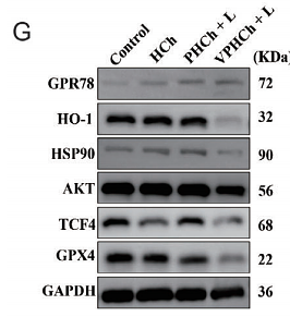
Tumor Microenvironment-Driven Structural Transformation of Vanadium-Based MXenzymes to Amplify Oxidative Stress for Multimodal Tumor Therapy
Author: Hai Zhu, Tinghua Li, Xinhao Peng, Xiaoxian Zhang, Xuequan Zhang, Qiusheng Wang, Lei Lei, Jun Zhang, Bin He, Jun Cao
PMID: 39853632
Journal: Advanced Science
Application: WB
Reactivity: Mouse
Publish date: 2025 Jan
-
Citation
-
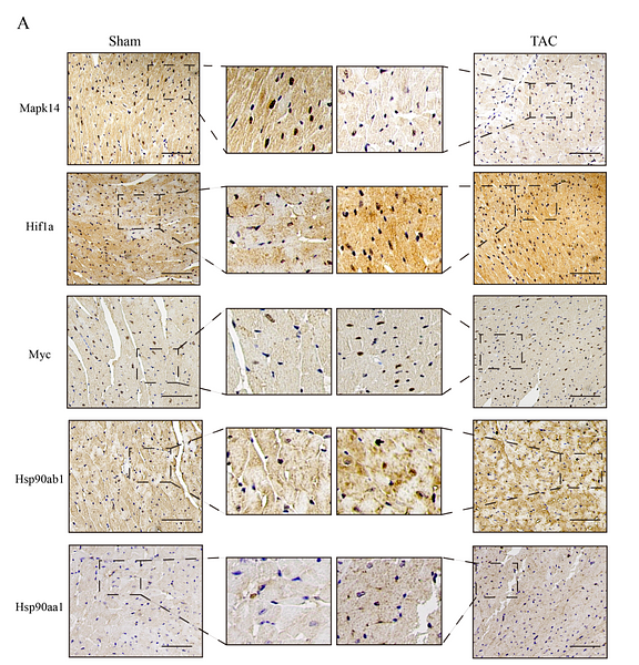
Identification of hub genes associated with oxidative stress in heart failure and their correlation with immune infiltration using bioinformatics analysis
Author: Jianjun Gu, Li Na Zhang, Xiang Gu, Ye Zhu
PMID: 37609434
Journal: Peer J
Application: IHC
Reactivity: Mouse
Publish date: 2023 Aug
-
Citation
-
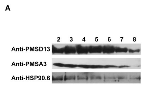
HEAT SHOCK PROTEIN 90.6 interacts with carbon and nitrogen metabolism components during seed development
Author:
PMID: 36652388
Journal: Plant Physiology
Application: WB
Reactivity: Zea mays
Publish date: 2023 Apr
-
Citation
-
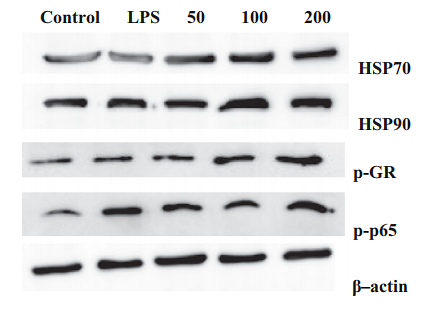
Amelioration of LPS-induced inflammatory response and oxidative stress by astaxanthin in Channa argus lymphocyte via activating glucocorticoid receptor signalling pathways
Author: Mu-Yang Li, Chun-Shan Gao, Xiao-Yan Du, Lei Zhao, Xiao-Tian Niu, Gui-Qin Wang, Dong-Ming Zhang
PMID: 3NOPMID25021001
Journal: Aquaculture Research
Application:
Reactivity:
Publish date: 2020 Mar
-
Citation
Products with the same target and pathway
HSP90 Beta Mouse Monoclonal Antibody [2-1-G3]
Application: WB,IHC-P
Reactivity: Human,Mouse,Rat
Conjugate: unconjugated
HSP90 Beta Recombinant Mouse Monoclonal Antibody [2-1-G3-R]
Application: WB,IHC-P,IF-Cell
Reactivity: Human,Mouse,Rat
Conjugate: unconjugated