
HRP Conjugated beta Tubulin Recombinant Rabbit Monoclonal Antibody [SR25-04] - Loading control



Catalog# HA724236H
HRP Conjugated beta Tubulin Recombinant Rabbit Monoclonal Antibody [SR25-04] - Loading control
-
WB
-
Human
-
Mouse
-
Rat
-
Green monkey
-
HRP
Overview
Product Name
HRP Conjugated beta Tubulin Recombinant Rabbit Monoclonal Antibody [SR25-04] - Loading control
Antibody Type
Recombinant Rabbit monoclonal Antibody
Immunogen
Synthetic peptide within Human beta Tubulin aa 308-357 / 444.
Species Reactivity
Human, Mouse, Rat, Green monkey
Validated Applications
WB
Target Molecular Weight
Predicted band size: 50 kDa
Positive Control
HeLa cell lysate, HEK-293 cell lysate, NIH/3T3 cell lysate, RAW264.7 cell lysate, C6 cell lysate, PC-12 cell lysate, COS-1 cell lysate.
Conjugation
HRP
Clone Number
SR25-04
Product Features
Form
Liquid
Concentration
Storage Instructions
Shipped at 4℃. Store at +4℃ short term (1-2 weeks). Store at -20℃ long term.
Storage Buffer
1*PBS (pH7.4), 0.1% BSA, 40% Glycerol, 0.2% Proclean 950.
Isotype
IgG
Purification Method
Protein A affinity purified.
Application Dilution
-
WB
-
1:5,000-1:20,000
Target
Function
Tubulin is one of several members of a small family of globular proteins. The most common members of the tubulin family are α-tubulin and β-tubulin. The beta-tubulin (relative molecular weight about 50 kDa) is counterpart of alpha-tubulin in tubulin heterodimer, it is coded by multiple tubulin genes and it is also posttranslationally modified. Heterogeneity of subunit is concentrated in C-terminal structural domain. Beta-Tubulin may have bound GTP or GDP. Under certain conditions β-tubulin can hydrolyze its bound GTP to GDP plus Pi, release the Pi, and exchange the GDP for GTP.
Background References
1. "Tumoral and tissue-specific expression of the major human beta-tubulin isotypes."Leandro-Garcia L.J., Leskela S., Landa I., Montero-Conde C., Lopez-Jimenez E., Leton R., .Cytoskeleton 67:214-223(2010).
2. "Five mouse tubulin isotypes and their regulated expression during development."Lewis S.A., Lee M.G.-S., Cowan N.J.J. Cell Biol. 101:852-861(1985).
Sequence Similarity
Belongs to the tubulin family.
Tissue Specificity
Ubiquitously expressed with highest levels in spleen, thymus and immature brain.
Post-translational Modification
Some glutamate residues at the C-terminus are polyglutamylated, resulting in polyglutamate chains on the gamma-carboxyl group. Polyglutamylation plays a key role in microtubule severing by spastin (SPAST). SPAST preferentially recognizes and acts on microtubules decorated with short polyglutamate tails: severing activity by SPAST increases as the number of glutamates per tubulin rises from one to eight, but decreases beyond this glutamylation threshold.; Some glutamate residues at the C-terminus are monoglycylated but not polyglycylated due to the absence of functional TTLL10 in human. Monoglycylation is mainly limited to tubulin incorporated into axonemes (cilia and flagella). Both polyglutamylation and monoglycylation can coexist on the same protein on adjacent residues, and lowering glycylation levels increases polyglutamylation, and reciprocally. The precise function of monoglycylation is still unclear (Probable).; Phosphorylated on Ser-172 by CDK1 during the cell cycle, from metaphase to telophase, but not in interphase. This phosphorylation inhibits tubulin incorporation into microtubules.
Subcellular Location
Cytoplasm
Synonyms
Beta 4 tubulin antibody
Beta 5 tubulin antibody
BetaTubulin antibody
TBB5_HUMAN antibody
TUBB antibody
TUBB2 antibody
TUBB2A antibody
TUBB5 antibody
tubulin beta 2A antibody
Tubulin beta chain antibody
ExpandImages
-
Western blot analysis of beta Tubulin on different lysates with Mouse anti-beta Tubulin antibody (HA724236H) at 1/20,000 dilution.
Lane 1: HeLa cell lysate
Lane 2: HEK-293 cell lysate
Lane 3: NIH/3T3 cell lysate
Lane 4: RAW264.7 cell lysate
Lane 5: C6 cell lysate
Lane 6: PC-12 cell lysate
Lane 7: COS-1 cell lysate
Lysates/proteins at 10 µg/Lane.
Predicted band size: 50 kDa
Observed band size: 50 kDa
Exposure time: 54 seconds; ECL: K1801;
4-20% SDS-PAGE gel.
Proteins were transferred to a PVDF membrane and blocked with 5% NFDM/TBST for 1 hour at room temperature. The primary antibody (HA724236H, HRP) at 1/20,000 dilution was used in primary antibody dilution buffer (K1803) at room temperature for 1 hour.
Please note: All products are "FOR RESEARCH USE ONLY AND ARE NOT INTENDED FOR DIAGNOSTIC OR THERAPEUTIC USE"
Products with the same target and pathway

beta Tubulin Recombinant Rabbit Monoclonal Antibody [SR25-04] - BSA and Azide free
Application: WB,IF-Cell,IHC-P,FC
Reactivity: Human,Mouse,Rat
Conjugate: unconjugated

beta Tubulin Mouse Monoclonal Antibody [1-B11]
Application: WB,IF-Cell,IHC-P,FC,IF-Tissue
Reactivity: Human,Mouse,Rat,Zebrafish
Conjugate: unconjugated

beta Tubulin Mouse Monoclonal Antibody [A1-A4]
Application: WB,IF-Cell,IHC-P,FC
Reactivity: Human,Mouse,Rat,Green monkey
Conjugate: unconjugated

beta Tubulin Recombinant Mouse Monoclonal Antibody [A1-A4-R] - BSA and Azide free
Application: WB,IF-Cell,FC,IHC-P
Reactivity: Human,Mouse,Rat,Green monkey
Conjugate: unconjugated

beta Tubulin Recombinant Mouse Monoclonal Antibody [1-B11-R]
Application: WB,IF-Cell,IHC-P
Reactivity: Human,Mouse,Rat
Conjugate: unconjugated
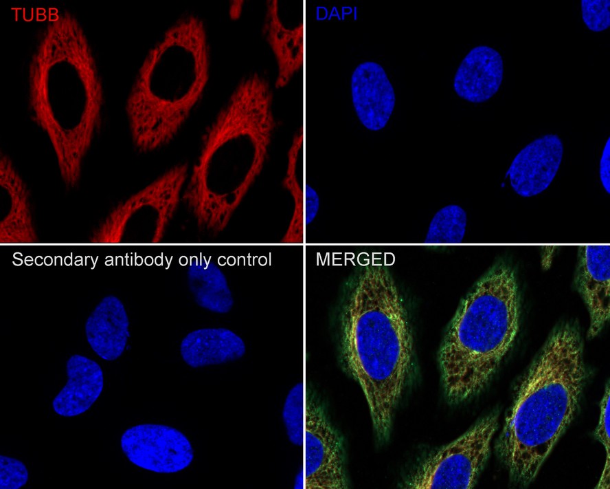
iFluor™ 594 Conjugated beta Tubulin Recombinant Mouse Monoclonal Antibody [A1-A4-R]
Application: IF-Cell,IF-Tissue,FC
Reactivity: Human,Mouse
Conjugate: iFluor™ 594

beta Tubulin Recombinant Mouse Monoclonal Antibody [1-B11-R] - BSA and Azide free
Application: WB,IF-Cell,IHC-P
Reactivity: Human,Mouse,Rat
Conjugate: unconjugated

beta Tubulin Recombinant Rabbit Monoclonal Antibody [SR25-04]
Application: WB,IF-Cell,IHC-P,FC
Reactivity: Human,Mouse,Rat
Conjugate: unconjugated

HRP Conjugated beta Tubulin Mouse Monoclonal Antibody [1-B11]
Application: WB,IHC-P
Reactivity: Human,Mouse,Rat,Zebrafish
Conjugate: HRP

HRP Conjugated beta Tubulin Recombinant Rabbit Monoclonal Antibody [JF41-50]
Application: WB
Reactivity: Human,Mouse,Rat
Conjugate: HRP
