Specification
Catalog# ET1609-25
TOMM20 Recombinant Rabbit Monoclonal Antibody [ST04-72]
-
WB
-
IHC-P
-
IF-Tissue
-
IF-Cell
-
FC
-
IP
-
IHC-Fr
-
Human
-
Mouse
-
Rat
-
HA750166
不含抗保成分
-
ET1609-25
含抗保成分
-
unconjugated
Overview
Product Name
TOMM20 Recombinant Rabbit Monoclonal Antibody [ST04-72]
Antibody Type
Recombinant Rabbit monoclonal Antibody
Immunogen
Recombinant protein within Human TOMM20 aa 1-145 / 145.
Species Reactivity
Human, Mouse, Rat
Validated Applications
WB, IHC-P, IF-Tissue, IF-Cell, FC, IP, IHC-Fr
Molecular Weight
Predicted band size: 16 kDa
Positive Control
HeLa cell lysate, Saos-2 cell lysate, HepG2 cell lysate, A549 cell lysate, NIH/3T3 cell lysate, C2C12 cell lysate, C6 cell lysate, PC-12 cell lysate, Mouse brain tissue lysate, Rat brain tissue lysate, MCF7 cell lysate, F9 cell lysate, Rat lung tissue lysate, HepG2, NIH/3T3, HeLa, human kidney tissue, rat kidney tissue, mouse kidney tissue, human liver tissue, rat large intestine tissue.
Conjugation
unconjugated
Clone Number
ST04-72
RRID
Product Features
Form
Liquid
Storage Instructions
Shipped at 4℃. Store at +4℃ short term (1-2 weeks). It is recommended to aliquot into single-use upon delivery. Store at -20℃ long term.
Storage Buffer
1*TBS (pH7.4), 0.05% BSA, 40% Glycerol. Preservative: 0.05% Sodium Azide.
Isotype
IgG
Purification Method
Protein A affinity purified.
Application Dilution
-
WB
-
1:5,000-1:20,000
-
IHC-P
-
1:500-1:1,000
-
IHC-Fr
-
1:500
-
IF-Tissue
-
1:500
-
IF-Cell
-
1:1,000
-
FC
-
1:1,000
-
IP
-
Use at an assay dependent concentration.
Applications in Publications
Species in Publications
| Mouse | See 15 publications below |
| Human | See 7 publications below |
| Chicken | See 3 publications below |
| Fish | See 1 publications below |
| Grass carp | See 1 publications below |
| Rat | See 1 publications below |
| Hen | See 1 publications below |
Target
Function
Mitochondrial import receptor subunit TOM20 homolog is a protein that in humans is encoded by the TOMM20 gene. TOM20 is one of the receptor systems of the translocase of the outer membrane (TOM) complex in the outer mitochondrial membrane. In mitochondrial protein import, TOM20 is closely associated with the pore-forming TOM40 complex and acts by recognizing and binding the N-terminal MTSs (matrix-targeting sequences), which form an amphipathic alpha helix and aid passage of the target proteins into the mitochondrial matrix.
Background References
1. Kim SJ et al. Hepatitis C virus induces the mitochondrial translocation of Parkin and subsequent mitophagy. PLoS Pathog 9:e1003285 (2013).
2. Kim SJ et al. Hepatitis B virus disrupts mitochondrial dynamics: induces fission and mitophagy to attenuate apoptosis. PLoS Pathog 9:e1003722 (2013).
Sequence Similarity
Belongs to the Tom20 family.
Post-translational Modification
Ubiquitinated by PRKN during mitophagy, leading to its degradation and enhancement of mitophagy. Deubiquitinated by USP30.
Subcellular Location
Mitochondrion outer membrane.
Synonyms
KIAA0016 antibody
MAS20 antibody
MGC117367 antibody
Mitochondrial 20 kDa outer membrane protein antibody
Mitochondrial import receptor subunit TOM20 homolog antibody
MOM19 antibody
Outer mitochondrial membrane receptor Tom20 antibody
TOM20 antibody
TOM20_HUMAN antibody
TOMM20 antibody
ExpandKIAA0016 antibody
MAS20 antibody
MGC117367 antibody
Mitochondrial 20 kDa outer membrane protein antibody
Mitochondrial import receptor subunit TOM20 homolog antibody
MOM19 antibody
Outer mitochondrial membrane receptor Tom20 antibody
TOM20 antibody
TOM20_HUMAN antibody
TOMM20 antibody
Translocase of outer mitochondrial membrane 20 homolog (yeast) antibody
Translocase of outer mitochondrial membrane 20 homolog type II antibody
CollapseImages
-

☑ Relative expression (RE)
Western blot analysis of TOMM20 on different lysates with Rabbit anti-TOMM20 antibody (ET1609-25) at 1/5,000 dilution and competitor's antibody at 1/1,000 dilution.
Lane 1: HeLa cell lysate (15 µg/Lane)
Lane 2: Saos-2 cell lysate (low expression) (15 µg/Lane)
Lane 3: HepG2 cell lysate (15 µg/Lane)
Lane 4: A549 cell lysate (15 µg/Lane)
Lane 5: NIH/3T3 cell lysate (15 µg/Lane)
Lane 6: C2C12 cell lysate (15 µg/Lane)
Lane 7: C6 cell lysate (15 µg/Lane)
Lane 8: PC-12 cell lysate (15 µg/Lane)
Lane 9: Mouse brain tissue lysate (30 µg/Lane)
Lane 10: Rat brain tissue lysate (30 µg/Lane)
Predicted band size: 16 kDa
Observed band size: 16 kDa
Exposure time: 3 minutes; ECL: K1801;
4-20% SDS-PAGE gel.
Proteins were transferred to a PVDF membrane and blocked with 5% NFDM/TBST for 1 hour at room temperature. The primary antibody (ET1609-25) at 1/5,000 dilution and competitor's antibody at 1/1,000 dilution were used in 5% NFDM/TBST at 4℃ overnight. Goat Anti-Rabbit IgG - HRP Secondary Antibody (HA1001) at 1/50,000 dilution was used for 1 hour at room temperature. -

☑ Knockdown (KD)
Western blot analysis of TOMM20 on different lysates with Rabbit anti-TOMM20 antibody (ET1609-25) at 1/5,000 dilution.
Lane 1: HeLa-si NT cell lysate (10 µg/Lane)
Lane 2: HeLa-si TOMM20 cell lysate (10 µg/Lane)
Predicted band size: 16 kDa
Observed band size: 16 kDa
Exposure time: 21 seconds; ECL: K1801;
4-20% SDS-PAGE gel.
Proteins were transferred to a PVDF membrane and blocked with 5% NFDM/TBST for 1 hour at room temperature. The primary antibody (ET1609-25) at 1/5,000 dilution was used in 5% NFDM/TBST at 4℃ overnight. Goat Anti-Rabbit IgG - HRP Secondary Antibody (HA1001) at 1/50,000 dilution was used for 1 hour at room temperature. -

☑ Relative expression (RE)
Western blot analysis of TOMM20 on different lysates with Rabbit anti-TOMM20 antibody (ET1609-25) at 1/5,000 dilution.
Lane 1: HeLa cell lysate (10 µg/Lane)
Lane 2: Saos-2 cell lysate (low expression) (10 µg/Lane)
Lane 3: HepG2 cell lysate (10 µg/Lane)
Lane 4: A549 cell lysate (10 µg/Lane)
Lane 5: MCF7 cell lysate (10 µg/Lane)
Lane 6: NIH/3T3 cell lysate (10 µg/Lane)
Lane 7: F9 cell lysate (10 µg/Lane)
Lane 8: PC-12 cell lysate (10 µg/Lane)
Lane 9: Mouse brain tissue lysate (20 µg/Lane)
Lane 10: Rat brain tissue lysate (20 µg/Lane)
Lane 11: Rat lung tissue lysate (20 µg/Lane)
Predicted band size: 16 kDa
Observed band size: 16 kDa
Exposure time: 1 minute 22 seconds; ECL: K1801;
4-20% SDS-PAGE gel.
Proteins were transferred to a PVDF membrane and blocked with 5% NFDM/TBST for 1 hour at room temperature. The primary antibody (ET1609-25) at 1/5,000 dilution was used in 5% NFDM/TBST at room temperature for 2 hours. Goat Anti-Rabbit IgG - HRP Secondary Antibody (HA1001) at 1/100,000 dilution was used for 1 hour at room temperature. -

Application: IF-cell
Species: Human
Cell Sample: HeLa cell
Antibody concentration: 1: 1,000
Date by conrtesy of: Mr. Wenxiang Huang
School of Basic Medical Sicences, Zhejiang University -

Application: IF-cell
Species: Human
Cell Sample: AC16 cell
Antibody concentration: 1: 200
Date by conrtesy of: Mr. Zhiyi Yang
School of Basic Medical Sicences, Zhejiang University -

☑ Relative expression (RE)
Immunocytochemistry analysis of HepG2 (high expression) and Saos-2 (low expression) labeling TOMM20 with Rabbit anti-TOMM20 antibody (ET1609-25) at 1/1,000 dilution and competitor's antibody at 1/400 dilution.
Cells were fixed in 4% paraformaldehyde for 20 minutes at room temperature, permeabilized with 0.1% Triton X-100 in PBS for 5 minutes at room temperature, then blocked with 1% BSA in 10% negative goat serum for 1 hour at room temperature. Cells were then incubated with Rabbit anti-TOMM20 antibody (ET1609-25) at 1/1,000 dilution and competitor's antibody at 1/400 dilution in 1% BSA in PBST overnight at 4 ℃. Goat Anti-Rabbit IgG H&L (iFluor™ 488, HA1121) was used as the secondary antibody at 1/1,000 dilution. PBS instead of the primary antibody was used as the secondary antibody only control. Nuclear DNA was labelled in blue with DAPI.
Beta tubulin (M1305-2, red) was stained at 1/100 dilution overnight at +4℃. Goat Anti-Mouse IgG H&L (iFluor™ 594, HA1126) was used as the secondary antibody at 1/1,000 dilution. -

Immunocytochemistry analysis of NIH/3T3 cells labeling TOMM20 with Rabbit anti-TOMM20 antibody (ET1609-25) at 1/1,000 dilution and competitor's antibody at 1/400 dilution.
Cells were fixed in 4% paraformaldehyde for 20 minutes at room temperature, permeabilized with 0.1% Triton X-100 in PBS for 5 minutes at room temperature, then blocked with 1% BSA in 10% negative goat serum for 1 hour at room temperature. Cells were then incubated with Rabbit anti-TOMM20 antibody (ET1609-25) at 1/1,000 dilution and competitor's antibody at 1/400 dilution in 1% BSA in PBST overnight at 4 ℃. Goat Anti-Rabbit IgG H&L (iFluor™ 488, HA1121) was used as the secondary antibody at 1/1,000 dilution. PBS instead of the primary antibody was used as the secondary antibody only control. Nuclear DNA was labelled in blue with DAPI.
Beta tubulin (M1305-2, red) was stained at 1/100 dilution overnight at +4℃. Goat Anti-Mouse IgG H&L (iFluor™ 594, HA1126) was used as the secondary antibody at 1/1,000 dilution. -

Immunocytochemistry analysis of HeLa cells labeling TOMM20 with Rabbit anti-TOMM20 antibody (ET1609-25) at 1/1,000 dilution.
Cells were fixed in 4% paraformaldehyde for 15 minutes at room temperature, permeabilized with 0.1% Triton X-100 in PBS for 15 minutes at room temperature, then blocked with 1% BSA in 10% negative goat serum for 1 hour at room temperature. Cells were then incubated with Rabbit anti-TOMM20 antibody (ET1609-25) at 1/1,000 dilution in 1% BSA in PBST overnight at 4 ℃. Goat Anti-Rabbit IgG H&L (iFluor™ 488, HA1121) was used as the secondary antibody at 1/1,000 dilution. PBS instead of the primary antibody was used as the secondary antibody only control. Counterstained with Mitotracker. Nuclear DNA was labelled in blue with DAPI. -

Immunohistochemical analysis of paraffin-embedded human kidney tissue with Rabbit anti-TOMM20 antibody (ET1609-25) at 1/500 dilution.
The section was pre-treated using heat mediated antigen retrieval with Tris-EDTA buffer (pH 9.0) for 20 minutes. The tissues were blocked in 1% BSA for 20 minutes at room temperature, washed with ddH2O and PBS, and then probed with the primary antibody (ET1609-25) at 1/500 dilution for 1 hour at room temperature. The detection was performed using an HRP conjugated compact polymer system. DAB was used as the chromogen. Tissues were counterstained with hematoxylin and mounted with DPX. -

Immunohistochemical analysis of paraffin-embedded rat kidney tissue with Rabbit anti-TOMM20 antibody (ET1609-25) at 1/500 dilution.
The section was pre-treated using heat mediated antigen retrieval with Tris-EDTA buffer (pH 9.0) for 20 minutes. The tissues were blocked in 1% BSA for 20 minutes at room temperature, washed with ddH2O and PBS, and then probed with the primary antibody (ET1609-25) at 1/500 dilution for 1 hour at room temperature. The detection was performed using an HRP conjugated compact polymer system. DAB was used as the chromogen. Tissues were counterstained with hematoxylin and mounted with DPX. -

Immunohistochemical analysis of paraffin-embedded mouse kidney tissue with Rabbit anti-TOMM20 antibody (ET1609-25) at 1/500 dilution.
The section was pre-treated using heat mediated antigen retrieval with Tris-EDTA buffer (pH 9.0) for 20 minutes. The tissues were blocked in 1% BSA for 20 minutes at room temperature, washed with ddH2O and PBS, and then probed with the primary antibody (ET1609-25) at 1/500 dilution for 1 hour at room temperature. The detection was performed using an HRP conjugated compact polymer system. DAB was used as the chromogen. Tissues were counterstained with hematoxylin and mounted with DPX. -

Immunohistochemical analysis of paraffin-embedded human liver tissue with Rabbit anti-TOMM20 antibody (ET1609-25) at 1/800 dilution.
The section was pre-treated using heat mediated antigen retrieval with Tris-EDTA buffer (pH 9.0) for 20 minutes. The tissues were blocked in 1% BSA for 20 minutes at room temperature, washed with ddH2O and PBS, and then probed with the primary antibody (ET1609-25) at 1/800 dilution for 1 hour at room temperature. The detection was performed using an HRP conjugated compact polymer system. DAB was used as the chromogen. Tissues were counterstained with hematoxylin and mounted with DPX. -

Immunohistochemical analysis of paraffin-embedded rat large intestine tissue with Rabbit anti-TOMM20 antibody (ET1609-25) at 1/800 dilution.
The section was pre-treated using heat mediated antigen retrieval with Tris-EDTA buffer (pH 9.0) for 20 minutes. The tissues were blocked in 1% BSA for 20 minutes at room temperature, washed with ddH2O and PBS, and then probed with the primary antibody (ET1609-25) at 1/800 dilution for 1 hour at room temperature. The detection was performed using an HRP conjugated compact polymer system. DAB was used as the chromogen. Tissues were counterstained with hematoxylin and mounted with DPX. -

Application: IHC-Fr
Species: Mouse
Site: Kidney
Sample: Frozen section
Antibody concentration: 1/500
Antigen retrieval: Not required -

Application: IHC-Fr
Species: Rat
Site: Kidney
Sample: Frozen section
Antibody concentration: 1/500
Antigen retrieval: Not required -

TOMM20 was immunoprecipitated in 0.2mg HeLa cell lysate with ET1609-25 at 2 µg/10 µl beads. Western blot was performed from the immunoprecipitate using ET1609-25 at 1/1,000 dilution. Anti-Rabbit IgG for IP Nano-secondary antibody (NBI01H) at 1/5,000 dilution was used for 1 hour at room temperature.
Lane 1: HeLa cell lysate (input)
Lane 2: Rabbit IgG instead of ET1609-25 in HeLa cell lysate
Lane 3: ET1609-25 IP in HeLa cell lysate
Blocking/Dilution buffer: 5% NFDM/TBST
Exposure time: 1 minute 2 seconds -

Flow cytometric analysis of HeLa cells labeling TOMM20.
Cells were fixed and permeabilized. Then stained with the primary antibody (ET1609-25, 1μg/mL) (red) compared with Rabbit IgG Isotype Control (green). After incubation of the primary antibody at +4℃ for an hour, the cells were stained with a iFluor™ 488 conjugate-Goat anti-Rabbit IgG Secondary antibody (HA1121) at 1/1,000 dilution for 30 minutes at +4℃. Unlabelled sample was used as a control (cells without incubation with primary antibody; black).
Please note: All products are "FOR RESEARCH USE ONLY AND ARE NOT INTENDED FOR DIAGNOSTIC OR THERAPEUTIC USE"
Citation
-
Preventive Effect of Fisetin on Follicular Granulosa Cells Senescence via Attenuating Oxidative Stress and Upregulating the Wnt/β-Catenin Signaling Pathway
Author: Juan Dong, Zhaoyu Yang, Qiongyu Yuan, Weidong Zeng, Yuling Mi, Caiqiao Zhang
PMID: 41227350
Journal: Cells
Application: WB
Reactivity: Hen
Publish date: 2025 Oct
-
Citation
-
tRNA-derived fragment tRF-24 drives CELF1 phase separation to promote oncogenic splicing in esophageal squamous cell carcinoma
Author: Hu Yajie, Qin Xin, Gong Li, Pan Ling, Cheng Yufeng
PMID: 10.1186/s13046-025-03553-x
Journal: Journal Of Experimental & Clinical Cancer Research
Application: WB
Reactivity: Human
Publish date: 2025 Nov
-
Citation
-
Targeted Delivery of AGO-2 to Myocardial Mitochondria via Functionalized Nanoparticles Attenuates Oxidative Stress in Diabetic Cardiomyopathy
Author: Wenbo Du, Yu Jiang, Lan Wu, Yuan Liang, Yu-Ping Wu, Hanrui Liu, Tian-Wu Chen
PMID: 10.1021/acsami.5c15154
Journal: ACS Applied Materials & Interfaces
Application: IF
Reactivity: Mouse
Publish date: 2025 Nov
-
Citation
-
The activation of cGAS-STING pathway promotes the epithelial-mesenchymal transition and inflammation in intrauterine adhesion
Author: Xixi Wu, Li He, Yonghong Lin, Yunfeng Zheng, Ran Mao, Jianguo Hu, Rui Yuan, Huisheng Ge
PMID: 40409098
Journal: International Immunopharmacology
Application: IF
Reactivity: Human
Publish date: 2025 May
-
Citation
-
Compression force regulates cementoblast mineralization via S1PR1/mitophagy axis
Author: Han Wang, Jingwen Cai, Linxin Chen, Sihang Chen, Xinhan Yang, Zhonghan Chen, Linyu Xu
PMID: 40035536
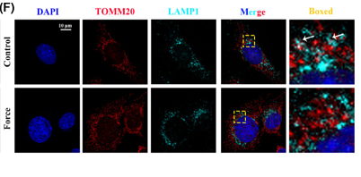
Journal: FASEB Journal
Application: IF
Reactivity: Human
Publish date: 2025 Mar
-
Citation
-
Impact of exosomes derived from adipose stem cells on lymphocyte proliferation and phenotype in mouse skin grafts
Author:
PMID: 10.20517/evcna.2024.52
Journal: Extracellular Vesicles and Circulating Nucleic Acids
Application: WB
Reactivity: Mouse
Publish date: 2025 Mar
-
Citation
-
Beta-asarone alleviated cerebral ischemia/reperfusion injury by targeting PINK1/Parkin-dependent mitophagy
Author: Yujiao Wang, Daojun Xie, Shijia Ma, Yuhe Wang, Chengcheng Zhang, Zhuyue Chen
PMID: 40490171
Journal: European Journal Of Pharmacology
Application: IF
Reactivity: Rat
Publish date: 2025 Jun
-
Citation
-
XBP1 promotes endometrial fibrosis through cGAS-STING signaling pathway in intrauterine adhesion
Author: Wu Xixi, He Li, Lin Yonghong, Zheng Yunfeng, Jiang Peng, Tian Chenfan, Mao Ran, Yang Bo, Shi Yuanling, Ge Huisheng, Hu Jianguo, Yuan Rui
PMID: 40603996
Journal: Scientific Reports
Application: IF
Reactivity: Human
Publish date: 2025 Jul
-
Citation
-
Kindlin-1 promotes mitophagy by inhibiting PINK1 degradation to enhance hepatocellular carcinoma progression and modulates sensitivity to donafenib
Author: Huaxing Ma, Guangling Ou, Bibo Wu, Hongwei Ding, Yijie Zhang, Fei Xia, Zixuan Shen, Kunyang Zhao, Chaochun Chen, Long Wu, Jin Lei, Yuan Xu, Xueke Zhao, Kun Cao, Haiyang Li
PMID: 40744334
Journal: Cellular Signalling
Application: WB
Reactivity: Human
Publish date: 2025 Jul
-
Citation
-
Macrophages and macrophage extracellular vesicles confer cancer ferroptosis resistance via PRDX6-mediated mitophagy inhibition
Author: Naisheng Zheng, Fuli Li, Qing Huang, Xian Huang, Tomasz Maj
PMID: 40825268
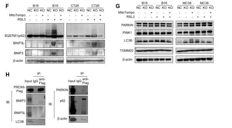
Journal: Redox Biology
Application: WB
Reactivity: Mouse
Publish date: 2025 Aug
-
Citation
-
The NR3C2-SIRT1 signaling axis promotes autophagy and inhibits epithelial mesenchymal transition in colorectal cancer
Author:
PMID: 40229278
Journal:
Application: WB
Reactivity: Mouse
Publish date: 2025 Apr
-
Citation
-
Integrated Analysis of PSMB8 Expression and Its Potential Roles in Hepatocellular Carcinoma
Author: Lu Ruijiao, Abuduhailili Xieyidai, Li Yuxia, Wang Senyu, Xia Xigang, Feng Yangchun
PMID: 40261568
Journal: Digestive Diseases and Sciences
Application: IF
Reactivity: Human
Publish date: 2025 Apr
-
Citation
-
Unconjugated bilirubin promotes uric acid restoration by activating hepatic AMPK pathway
Author: Yingqiong Zhang, Yujia Chen, Xiaojing Chen, Yue Gao, Jun Luo, Shuanghui Lu, Qi Li, Ping Li, Mengru Bai, Ting Jiang, Nanxin Zhang, Bichen Zhang, Binxin Chen, Hui Zhou, Huidi Jiang, Nengming Lin
PMID: 39299526
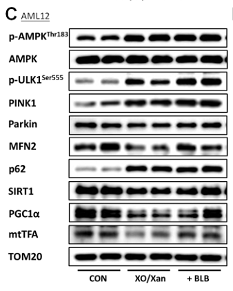
Journal: Free Radical Biology And Medicine
Application: WB
Reactivity: Mouse
Publish date: 2024 Sept
-
Citation
-
Sortilin-mediated translocation of mitochondrial ACSL1 impairs adipocyte thermogenesis and energy expenditure in male mice
Author: Min Yang,et al
PMID: 39232011
Journal: Nature Communications
Application: IHC-P
Reactivity: Mouse
Publish date: 2024 Sep
-
Citation
-
Edwardsiella piscicida promotes mitophagy to escape autophagy-mediated antibacterial defense in teleost monocytes/macrophages
Author: Jiaxi Liu,et al
PMID: NO PMID 2024102503
Journal: Aquaculture
Application: IF
Reactivity: Grass carp
Publish date: 2024 Oct
-
Citation
-
NDR2 is critical for the osteoclastogenesis by regulating ULK1-mediated mitophagy
Author: Xiangxi Kong, Zhi Shan, Yihao Zhao, Siyue Tao, Jingyun Chen, Zhongyin Ji, Jiayan Jin, Junhui Liu, Wenlong Lin, Xiaojian Wang, Jian Wang, Fengdong Zhao, Bao Huang, Jian Chen
PMID: 39561008
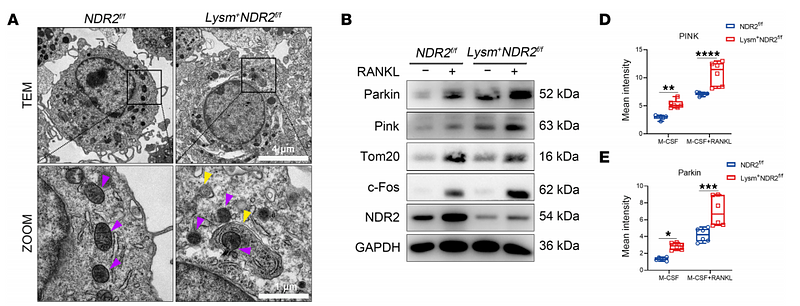
Journal: Journal Of Clinical Investigation Insight
Application: WB
Reactivity: Mouse
Publish date: 2024 Nov
-
Citation
-
NPR1 promotes cisplatin resistance by inhibiting PARL-mediated mitophagy-dependent ferroptosis in gastric cancer
Author: Chengwei Wu,et al
PMID: 39476297
Journal: Cell Biology And Toxicology
Application: WB
Reactivity: Mouse
Publish date: 2024 Nov
-
Citation
-
Loss of CHCHD2 Stability Coordinates with C1QBP/CHCHD2/CHCHD10 Complex Impairment to Mediate PD-Linked Mitochondrial Dysfunction
Author: Ren Yanlin,et al
PMID: 38453793
Journal: Molecular Neurobiology
Application: WB
Reactivity: Mouse
Publish date: 2024 Mar
-
Citation
-
Lactobacillus salivarius metabolite succinate enhances chicken intestinal stem cell activities via the SUCNR1-mitochondria axis
Author: Danni Luo, Minyao Zou, Xi Rao, Mingping Wei, Lingzhi Zhang, Yuping Hua, Lingzi Yu, Jiajia Cao, Jinyi Ye, Sichao Qi, Huanan Wang, Yuling Mi, Caiqiao Zhang, Jian Li
PMID: 39764876
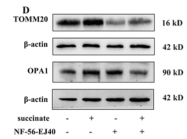
Journal: Poultry Science
Application: WB,IF
Reactivity: Chicken
Publish date: 2024 Dec
-
Citation
-
Krüppel-like factor 5 activates chick intestinal stem cell and promotes mucosal repair after impairment
Author: Yu L, Qi S, Wei G, et al
PMID: 37950881
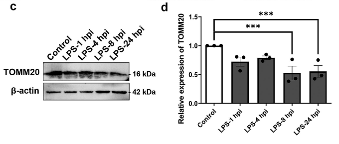
Journal: Cell Cycle
Application: WB
Reactivity: Chicken
Publish date: 2023 Nov
-
Citation
-
Wei-Tong-Xin ameliorated cisplatin-induced mitophagy and apoptosis in gastric antral mucosa by activating the Nrf2/HO-1 pathway
Author:
PMID: 36806345
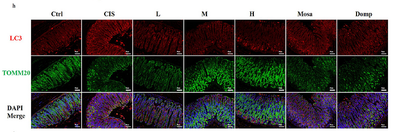
Journal: Journal Of Ethnopharmacology
Application: IF-tissue
Reactivity: Mouse
Publish date: 2023 May
-
Citation
-
Follicle-Stimulating Hormone Alleviates Ovarian Aging by Modulating Mitophagy- and Glycophagy-Based Energy Metabolism in Hens
Author: Dong, J., Guo, C., Yang, Z., Wu, Y., & Zhang, C.
PMID: 36291137
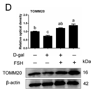
Journal: Cells
Application: WB,IF
Reactivity: Chicken
Publish date: 2022 Oct
-
Citation
-
SARS-CoV-2 ORF3a induces RETREG1/FAM134B dependent reticulophagy and triggers sequential ER stress and inflammatory responses during SARS-CoV-2 infection
Author: Zhang, X., Yang, Z., Pan, T., Long, X., Sun, Q., Wang, P. H., Li, X., & Kuang, E.
PMID: 35239449
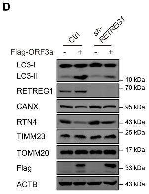
Journal: Autophagy
Application: WB
Reactivity: Human
Publish date: 2022 Mar
-
Citation
-
Physiological and transcriptomic analyses reveal the toxicological mechanism and risk assessment of environmentally-relevant waterborne tetracycline exposure on the gills of tilapia (Oreochromis niloticus)
Author:
PMID: 34743874
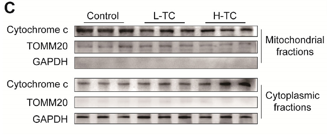
Journal: The Science Of The Total Environment
Application: WB
Reactivity: Fish
Publish date: 2022 Feb
-
Citation
-
Ubiquitination of NLRP3 by gp78/Insig-1 restrains NLRP3 inflammasome activation
Author: Xu, T., Yu, W., Fang, H., Wang, Z., Chi, Z., Guo, X., Jiang, D., Zhang, K., Chen, S., Li, M., Guo, Y., Zhang, J., Yang, D., Yu, Q., Wang, D., & Zhang, X.
PMID: 35110683
Journal: Cell Death & Differentiation
Application: WB
Reactivity: Mouse
Publish date: 2022 Feb
-
Citation
-
Epithelial Gasdermin D shapes the host-microbial interface by driving mucus layer formation
Author: Zhang, J., Yu, Q., Jiang, D., Yu, K., Yu, W., Chi, Z., Chen, S., Li, M., Yang, D., Wang, Z., Xu, T., Guo, X., Zhang, K., Fang, H., Ye, Q., He, Y., Zhang, X., & Wang, D.
PMID: 35119941
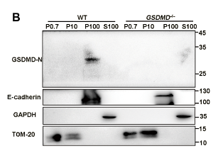
Journal: Science Immunology
Application: WB
Reactivity: Mouse
Publish date: 2022 Feb
-
Citation
-
Oxidative stress-mediated mitochondrial fission promotes hepatic stellate cell activation via stimulating oxidative phosphorylation
Author: Zhou, Y., Long, D., Zhao, Y., Li, S., Liang, Y., Wan, L., Zhang, J., Xue, F., & Feng, L.
PMID: 35933403
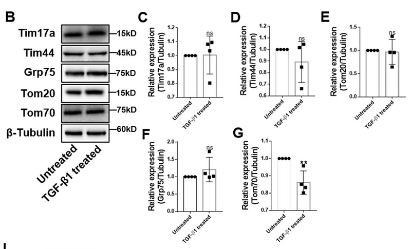
Journal: Cell Death & Disease
Application: WB
Reactivity: Mouse
Publish date: 2022 Aug
-
Citation
-
Autophagic elimination of ribosomes during spermiogenesis provides energy for flagellar motility
Author: Lei, Y., Zhang, X., Xu, Q., Liu, S., Li, C., Jiang, H., Lin, H., Kong, E., Liu, J., Qi, S., Li, H., Xu, W., & Lu, K.
PMID: 34428398
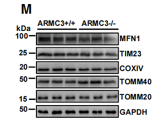
Journal: Developmental Cell
Application: WB
Reactivity: Mouse
Publish date: 2021 Aug
-
Citation
-
Histone Deacetylase 3 Couples Mitochondria to Drive IL-1β-Dependent Inflammation by Configuring Fatty Acid Oxidation
Author: Di Wang
PMID: 32937100
Journal: Molecular Cell
Application: WB,IP
Reactivity: Mouse
Publish date: 2020 Oct
-
Citation


