Specification
Catalog# ET1610-97
Hes1 Recombinant Rabbit Monoclonal Antibody [SC06-21]
-
WB
-
IHC-P
-
IHC-Fr
-
IF-Tissue
-
IF-Cell
-
FC
-
Human
-
Mouse
-
Rat
Overview
Product Name
Hes1 Recombinant Rabbit Monoclonal Antibody [SC06-21]
Antibody Type
Recombinant Rabbit monoclonal Antibody
Immunogen
Synthetic peptide within Human Hes1 aa 298-241 / 280.
Species Reactivity
Human, Mouse, Rat
Validated Applications
WB, IHC-P, IHC-Fr, IF-Tissue, IF-Cell, FC
Molecular Weight
Predicted band size: 30 kDa
Positive Control
SH-SY5Y cell lysate, MDA-MB-231 cell lysate, MCF7 cell lysate, SK-MEL-28 cell lysate, SH-SY5Y, mouse brain tissue, rat brain tissue, human breast carcinoma tissue, mouse cerebellum tissue.
Conjugation
unconjugated
Clone Number
SC06-21
RRID
Product Features
Form
Liquid
Concentration
1ug/ul
Storage Instructions
Shipped at 4℃. Store at +4℃ short term (1-2 weeks). It is recommended to aliquot into single-use upon delivery. Store at -20℃ long term.
Storage Buffer
1*TBS (pH7.4), 0.05% BSA, 40% Glycerol. Preservative: 0.05% Sodium Azide.
Isotype
IgG
Purification Method
Protein A affinity purified.
Application Dilution
-
WB
-
1:5,000
-
IHC-P
-
1:2,000-1:5,000
-
IHC-Fr
-
1:500
-
IF-Tissue
-
1:1,000
-
IF-Cell
-
1:2,000
-
FC
-
1:1,000
Applications in Publications
Species in Publications
Human | See 4 publications below |
Mouse | See 2 publications below |
Pig | See 1 publications below |
Target
Function
Transcription factor HES1 (hairy and enhancer of split-1) is a protein that is encoded by the Hes1 gene, and is the mammalian homolog of the hairy gene in Drosophila. HES1 is one of the seven members of the Hes gene family (HES1-7). Hes genes code nuclear proteins that suppress transcription. This protein belongs to the basic helix-loop-helix (bHLH) family of transcription factors. It is a transcriptional repressor of genes that require a bHLH protein for their transcription. The protein has a particular type of basic domain that contains a helix interrupting protein that binds to the N-box promoter region rather than the canonical enhancer box (E-box). As a member of the bHLH family, it is a transcriptional repressor that influences cell proliferation and differentiation in embryogenesis. HES1 regulates its own expression via a negative feedback loop, and oscillates with approximately 2-hour periodicity.
Background References
1. Liu W et al. MicroRNA-206 overexpression promotes apoptosis, induces cell cycle arrest and inhibits the migration of human hepatocellular carcinoma HepG2 cells. Int J Mol Med 34:420-8 (2014).
2. Tang Y et al. Rnd3 regulates lung cancer cell proliferation through notch signaling. PLoS One 9:e111897 (2014).
Subcellular Location
Nucleus.
Synonyms
bHLHb39 antibody
C-HAIRY1 antibody
c-hairy1A antibody
Class B basic helix-loop-helix protein 39 antibody
FLJ20408 antibody
Hairy and enhancer of split 1 (Drosophila) antibody
Hairy and enhancer of split 1 antibody
Hairy homolog (Drosophila) antibody
Hairy homolog antibody
Hairy like antibody
ExpandbHLHb39 antibody
C-HAIRY1 antibody
c-hairy1A antibody
Class B basic helix-loop-helix protein 39 antibody
FLJ20408 antibody
Hairy and enhancer of split 1 (Drosophila) antibody
Hairy and enhancer of split 1 antibody
Hairy homolog (Drosophila) antibody
Hairy homolog antibody
Hairy like antibody
Hairy, Drosophila, homolog of antibody
Hairy-like protein antibody
Hairy/enhancer of split, Drosophila, homolog of, 1 antibody
HAIRY1 antibody
HES-1 antibody
hes1 antibody
Hes1 hairy and enhancer of split 1 (Drosophila) antibody
HES1_HUMAN antibody
HHL antibody
HL antibody
HRY antibody
MGC129109 antibody
OTTHUMP00000209031 antibody
RHL antibody
Transcription factor HES-1 antibody
CollapseImages
-
Immunofluorescence analysis of frozen mouse cerebellum tissue with Rabbit anti-Hes1 antibody (ET1610-97) at 1/500 dilution.
The section was pre-treated using heat mediated antigen retrieval with sodium citrate buffer (pH 6.0) for about 2 minutes in microwave oven. The tissues were blocked in 10% negative goat serum for 1 hour at room temperature, washed with PBS, and then probed with the primary antibody (ET1610-97, green) at 1/500 dilution overnight at 4 ℃, washed with PBS. Goat Anti-Rabbit IgG H&L (iFluor™ 488, HA1121) was used as the secondary antibody at 1/1,000 dilution. Nuclei were counterstained with DAPI (blue). -
Western blot analysis of Hes1 on different lysates with Rabbit anti-Hes1 antibody (ET1610-97) at 1/5,000 dilution.
Lane 1: SH-SY5Y cell lysate
Lane 2: MDA-MB-231 cell lysate
Lane 3: MCF7 cell lysate
Lane 4: SK-MEL-28 cell lysate
Lysates/proteins at 15 µg/Lane.
Predicted band size: 30 kDa
Observed band size: 30 kDa
Exposure time: 1 minute 20 seconds;
4-20% SDS-PAGE gel.
Proteins were transferred to a PVDF membrane and blocked with 5% NFDM/TBST for 1 hour at room temperature. The primary antibody (ET1610-97) at 1/5,000 dilution was used in 5% NFDM/TBST at 4℃ overnight. Goat Anti-Rabbit IgG - HRP Secondary Antibody (HA1001) at 1:50,000 dilution was used for 1 hour at room temperature. -
Immunohistochemical analysis of paraffin-embedded mouse brain tissue with Rabbit anti-Hes1 antibody (ET1610-97) at 1/5,000 dilution.
The section was pre-treated using heat mediated antigen retrieval with Tris-EDTA buffer (pH 9.0) for 20 minutes. The tissues were blocked in 1% BSA for 20 minutes at room temperature, washed with ddH2O and PBS, and then probed with the primary antibody (ET1610-97) at 1/5,000 dilution for 1 hour at room temperature. The detection was performed using an HRP conjugated compact polymer system. DAB was used as the chromogen. Tissues were counterstained with hematoxylin and mounted with DPX. -
Immunohistochemical analysis of paraffin-embedded rat brain tissue with Rabbit anti-Hes1 antibody (ET1610-97) at 1/5,000 dilution.
The section was pre-treated using heat mediated antigen retrieval with Tris-EDTA buffer (pH 9.0) for 20 minutes. The tissues were blocked in 1% BSA for 20 minutes at room temperature, washed with ddH2O and PBS, and then probed with the primary antibody (ET1610-97) at 1/5,000 dilution for 1 hour at room temperature. The detection was performed using an HRP conjugated compact polymer system. DAB was used as the chromogen. Tissues were counterstained with hematoxylin and mounted with DPX. -
Immunohistochemical analysis of paraffin-embedded human breast cancer tissue with Rabbit anti-Hes1 antibody (ET1610-97) at 1/5,000 dilution.
The section was pre-treated using heat mediated antigen retrieval with Tris-EDTA buffer (pH 9.0) for 20 minutes. The tissues were blocked in 1% BSA for 20 minutes at room temperature, washed with ddH2O and PBS, and then probed with the primary antibody (ET1610-97) at 1/5,000 dilution for 1 hour at room temperature. The detection was performed using an HRP conjugated compact polymer system. DAB was used as the chromogen. Tissues were counterstained with hematoxylin and mounted with DPX. -
Immunofluorescence analysis of paraffin-embedded mouse brain tissue labeling Hes1 with Rabbit anti-Hes1 antibody (ET1610-97) at 1/1,000 dilution.
The section was pre-treated using heat mediated antigen retrieval with sodium citrate buffer (pH 6.0) (high pressure) for 2 minutes. The tissues were blocked in 10% negative goat serum for 1 hour at room temperature, washed with PBS, and then probed with the primary antibody (ET1610-97, green) at 1/1,000 dilution overnight at 4 ℃, washed with PBS.
Goat Anti-Rabbit IgG H&L (iFluor™ 488, HA1121) was used as the secondary antibody at 1/1,000 dilution. Nuclei were counterstained with DAPI (blue). -
Immunofluorescence analysis of paraffin-embedded rat brain tissue labeling Hes1 with Rabbit anti-Hes1 antibody (ET1610-97) at 1/500 dilution.
The section was pre-treated using heat mediated antigen retrieval with sodium citrate buffer (pH 6.0) (high pressure) for 2 minutes. The tissues were blocked in 10% negative goat serum for 1 hour at room temperature, washed with PBS, and then probed with the primary antibody (ET1610-97, green) at 1/500 dilution overnight at 4 ℃, washed with PBS.
Goat Anti-Rabbit IgG H&L (iFluor™ 488, HA1121) was used as the secondary antibody at 1/1,000 dilution. Nuclei were counterstained with DAPI (blue). -
Immunocytochemistry analysis of SH-SY5Y cells labeling Hes1 with Rabbit anti-Hes1 antibody (ET1610-97) at 1/2,000 dilution.
Cells were fixed in 4% paraformaldehyde for 20 minutes at room temperature, permeabilized with 0.1% Triton X-100 in PBS for 5 minutes at room temperature, then blocked with 1% BSA in 10% negative goat serum for 1 hour at room temperature. Cells were then incubated with Rabbit anti-Hes1 antibody (ET1610-97) at 1/2,000 dilution in 1% BSA in PBST overnight at 4 ℃. Goat Anti-Rabbit IgG H&L (iFluor™ 488, HA1121) was used as the secondary antibody at 1/1,000 dilution. PBS instead of the primary antibody was used as the secondary antibody only control. Nuclear DNA was labelled in blue with DAPI.
Beta tubulin (M1305-2, red) was stained at 1/100 dilution overnight at +4℃. Goat Anti-Mouse IgG H&L (iFluor™ 594, HA1126) was used as the secondary antibody at 1/1,000 dilution. -
Flow cytometric analysis of SH-SY5Y cells labeling Hes1.
Cells were fixed and permeabilized. Then stained with the primary antibody (ET1610-97, 1/1,000) (red) compared with Rabbit IgG Isotype Control (green). After incubation of the primary antibody at +4℃ for an hour, the cells were stained with a iFluor™ 488 conjugate-Goat anti-Rabbit IgG Secondary antibody (HA1121) at 1/1,000 dilution for 30 minutes at +4℃. Unlabelled sample was used as a control (cells without incubation with primary antibody; black).
Please note: All products are "FOR RESEARCH USE ONLY AND ARE NOT INTENDED FOR DIAGNOSTIC OR THERAPEUTIC USE"
Citation
-
Understanding the Role of NOTCH2 Mutation in Centronuclear Myopathy
Author: Youxi Lin, Hang Zhou, Wenjun Hu, Bo Gao, Tongzhou Liang, Jincheng Qiu, Pengfei Li, Yichen Que, Chipiu Wong, Xianjian Qiu, Zhihuai Deng, Huihong Shi, Song Liu, Jianan Chen, Nianchun Liao, Qihui Chen, Xiaojuan Li, Anjing Liang, Wenjie Gao, Dongsheng Huang
PMID: 40336196
Journal: Molecular Therapy
Application: WB
Reactivity: Mouse
Publish date: 2025 May
-
Citation
-
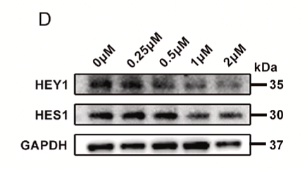
Taraxacum sinicum Kitag. (Binpu-3) root extract inhibits tumor invasion via Notch signaling in Drosophila and human breast cancer MDA-MB-231 cells
Author: Jiawei Wu, Jianbo Zhang, Wanyu Shu, Wei Feng, Ran Meng, Lingyu Kong, Huijuan Cao, Chunhua Jiang, Sitong Wang, Fanwu Wu, Chenxi Wu, Xiuping Wang
PMID: 40151788
Journal: Frontiers In Pharmacology
Application: WB
Reactivity: Human
Publish date: 2025 Mar
-
Citation
-
Notch signaling modulation enhances porcine muscle stem cell proliferation and differentiation
Author: Guanyu Qin, Zheng Liu, Hao Lu, Yumeng Zhang, Shijie Ding, Guanghong Zhou, Chunbao Li, Renpeng Guo
PMID: 39933473
Journal: Biochemical And Biophysical Research Communications
Application: WB
Reactivity: Pig
Publish date: 2025 Feb
-
Citation
-
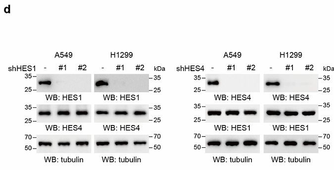
RNAi screens identify HES4 as a regulator of redox balance supporting pyrimidine synthesis and tumor growth
Author: He Jing,et al
PMID: 38769389
Journal: Nature Structural & Molecular Biology
Application: WB
Reactivity: Human
Publish date: 2024 May
-
Citation
-
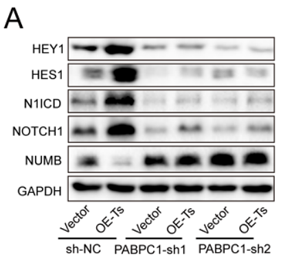
Exosomal long non-coding RNA TRPM2-AS promotes angiogenesis in gallbladder cancer through interacting with PABPC1 to activate NOTCH1 signaling pathway
Author: Zhiqiang He , Yuhan Zhong , Parbatraj Regmi , et al
PMID: 38532427
Journal: Molecular Cancer
Application: WB
Reactivity: Human
Publish date: 2024 Mar
-
Citation
-
Rho kinase inhibitor Y-27632 downregulates IL-1β expression in mice with experimental autoimmune myocarditis
Author: Li Yanjun, Gao Ge, Han Yiru, Xiao Bingshuai, Shen Liyuan, Yang Xiangxin, Liu Yangqing, Mu Yaqin, Zhang Nianping, Niu Chunhong, Wang Yuxing
PMID: 38684719
Journal: Scientific Reports
Application:
Reactivity:
Publish date: 2024 Apr
-
Citation
-
Jiawei Taohe Chengqi decoction inhibition of the notch signal pathway affects macrophage reprogramming to inhibit HSCs activation for the treatment of hepatic fibrosis
Author: Chang Shao, Huihui Xu, Xiguang Sun, Yun Pan, Xiaofan Liang, Jiaxin Huang, Yi He, Wenqin Guo, Linmao Ye, Junjie Zhang
PMID: 38030027
Journal: Journal Of Ethnopharmacology
Application: WB
Reactivity: Human
Publish date: 2023 Nov
-
Citation
-
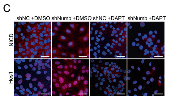
Loss of Numb promotes hepatic progenitor expansion and intrahepatic cholangiocarcinoma by enhancing Notch signaling
Author: Shu, Y., Xu, Q., Xu, Y., Tao, Q., Shao, M., Cao, X., Chen, Y., Wu, Z., Chen, M., Zhou, Y., Zhou, P., Shi, Y., & Bu, H.
PMID: 34667161
Journal: Cell Death & Disease
Application: WB,IHC-P,IF
Reactivity: Mouse
Publish date: 2021 Oct
-
Citation
Products with the same target and pathway
Hes1 Recombinant Rabbit Monoclonal Antibody [SC06-21] - BSA and Azide free
Application: WB,IHC-P,IHC-Fr,IF-Tissue,IF-Cell,FC
Reactivity: Human,Mouse,Rat
Conjugate: unconjugated