PD-L1 Recombinant Rabbit Monoclonal Antibody [PD01-02]
Specification
Catalog# HA721176
PD-L1 Recombinant Rabbit Monoclonal Antibody [PD01-02]
-
IHC-P
-
mIHC
-
WB
-
Human
-
unconjugated
Overview
Product Name
PD-L1 Recombinant Rabbit Monoclonal Antibody [PD01-02]
Antibody Type
Recombinant Rabbit monoclonal Antibody
Immunogen
Synthetic peptide within human PD-L1 aa 260-290 (Cytoplasmic).
Species Reactivity
Human
Validated Applications
IHC-P, mIHC, WB
Molecular Weight
Predicted band size: 33 kDa
Positive Control
Human Small Cell Lung Cancer, human non-small cell lung cancer, MDA-MB-231 cell lysate, U-87 MG cell lysate, HeLa cell lysate, A549 treated with 100ng/mL IFN gamma for 48 hours cell lysate, human lung carcinoma tissue, human placenta tissue.
Conjugation
unconjugated
Clone Number
PD01-02
RRID
Product Features
Form
Liquid
Storage Instructions
Shipped at 4℃. Store at +4℃ short term (1-2 weeks). It is recommended to aliquot into single-use upon delivery. Store at -20℃ long term.
Storage Buffer
PBS (pH7.4), 0.1% BSA, 40% Glycerol. Preservative: 0.05% Sodium Azide.
Isotype
IgG
Purification Method
Protein A affinity purified.
Application Dilution
-
IHC-P
-
1:200-1:800
-
mIHC
-
1:1,000
-
WB
-
1:2,000
Applications in Publications
Species in Publications
| Human | See 4 publications below |
| Mouse | See 1 publications below |
Target
Function
PD-L1 (programmed-death ligand 1; CD274), is a transmembrane protein constitutionally expressed on a variety of cell types, including antigen presenting cells (dendritic cells and histiocytes) and some non-lymphoid tissues (heart and lung). Binding of PD-L1 to PD-1 (programmed-death 1; CD279) expressed by activated T-cells, inhibits their function, causing negative feedback control of immunological reactions, thus impeding inflammation and autoimmunity. Tumour cells may express PD-L1, which binds to PD-1 allowing cancer cells to evade the attack of T-cells. Blockade of the PD-1/PD-L1 pathway has now shown useful in therapy of multiple cancer types, causing durable tumour regressions in a substantial proportion of otherwise treatment refractory cases of melanoma, and carcinomas of e.g., lung, kidney, and urinary tract. Patients without tumour PD-L1 expression can also derive benefit from blocking agents (studies across multiple cancer types demonstrate a pooled response rate of 48% in patients with PD-L1-positive tumours compared to 15% in PD-L1-negative tumours). Tonsil and placenta can be used as positive and negative tissue controls. However, tonsil is found to be superior to placenta, as tonsil displayes a range of PD-L1 expression levels. Tonsil displayes the following reaction pattern: No staining reaction in the vast majority of lymphocytes including mantle zone and germinal centre B-cells, no staining reaction in superficial epithelial cells, a weak to moderate, typically punctuated membranous staining reaction of the majority of germinal centre macrophages and finally a moderate to strong staining reaction of the majority of epithelial crypt cells.
Background References
1. Lei Q et al. Resistance Mechanisms of Anti-PD1/PDL1 Therapy in Solid Tumors. Front Cell Dev Biol. 2020 Jul
2. Tamene W et al. PDL1 expression on monocytes is associated with plasma cytokines in Tuberculosis and HIV. PLoS One. 2021 Oct
Subcellular Location
Cell membrane, Early endosome membrane, Recycling endosome membrane, Nucleus.
UNIPROT
Synonyms
B7 H antibody
B7 H1 antibody
B7 homolog 1 antibody
B7-H1 antibody
B7H antibody
B7H1 antibody
CD 274 antibody
CD-274 antibody
CD274 antibody
CD274 antigen antibody
ExpandB7 H antibody
B7 H1 antibody
B7 homolog 1 antibody
B7-H1 antibody
B7H antibody
B7H1 antibody
CD 274 antibody
CD-274 antibody
CD274 antibody
CD274 antigen antibody
CD274 molecule antibody
MGC142294 antibody
MGC142296 antibody
OTTHUMP00000021029 antibody
PD L1 antibody
PD-L1 antibody
PD1L1_HUMAN antibody
PDCD1 ligand 1 antibody
PDCD1L1 antibody
PDCD1LG1 antibody
PDL 1 antibody
PDL1 antibody
Programmed cell death 1 ligand 1 antibody
Programmed death ligand 1 antibody
RGD1566211 antibody
CollapseImages
-

Fluorescence multiplex immunohistochemical analysis of the human non-small cell lung cancer (Formalin/PFA-fixed paraffin-embedded sections). Panel A: the merged image of anti-CD20 (HA721138, green), anti-CD68 (HA601115, gray), anti-PD-L1 (HA721176, cyan), anti-panCK (HA601138, magenta) and anti-CD3 (HA720082, yellow) on human non-small cell lung cancer. Panel B: anti- CD20 stained on B cells. Panel C: anti-CD68 stained on macrophage M1 and macrophage M2. Panel D: anti-PD-L1 stained on dendritic cells and macrophages cells. Panel E: anti-panCK stained on cancer cells. Panel F: anti-CD3 stained on T cells. HRP Conjugated UltraPolymer Goat Polyclonal Antibody HA1119/HA1120 was used as a secondary antibody. The immunostaining was performed with the Sequential Immuno-staining Kit (IRISKit™MH010101, www.luminiris.cn). The section was incubated in five rounds of staining: in the order of HA721138 (1/1,500 dilution), HA601115 (1/2,000 dilution), HA721176 (1/1,000 dilution), HA601138 (1/3,000 dilution), and HA720082 (1/500 dilution) for 20 mins at room temperature. Each round was followed by a separate fluorescent tyramide signal amplification system. Heat mediated antigen retrieval with Tris-EDTA buffer (pH 9.0) for 30 mins at 95℃. DAPI (blue) was used as a nuclear counter stain. Image acquisition was performed with Olympus VS200 Slide Scanner.
-

Fluorescence multiplex immunohistochemical analysis of Tertiary Lymphoid Structures in Human Small Cell Lung Cancer (Formalin/PFA-fixed paraffin-embedded sections). Panel A: the merged image of anti-CD20 (HA721138, green), anti-PD-L1 (HA721176, cyan), anti-CD56 (ET1702-43, magenta) and anti-CD3 (HA720082, yellow) on tertiary lymphoid structures. Panel B: anti- CD20 stained on B cells. Panel C: anti-PD-L1 stained on dendritic cells and macrophages cells. Panel D: anti-CD56 stained on NKT cells. Panel E: anti-CD3 stained on T cells. HRP Conjugated UltraPolymer Goat Polyclonal Antibody HA1119/HA1120 was used as a secondary antibody. The immunostaining was performed with the Sequential Immuno-staining Kit (IRISKit™MH010101, www.luminiris.cn). The section was incubated in four rounds of staining: in the order of HA721138 (1/1,500 dilution), HA721176 (1/1,000 dilution), ET1702-43 (1/1,000 dilution), and HA720082 (1/500 dilution) for 20 mins at room temperature. Each round was followed by a separate fluorescent tyramide signal amplification system. Heat mediated antigen retrieval with Tris-EDTA buffer (pH 9.0) for 30 mins at 95℃. DAPI (blue) was used as a nuclear counter stain. Image acquisition was performed with Olympus VS200 Slide Scanner.
-

Fluorescence multiplex immunohistochemical analysis of Human non-small cell lung cancer (Formalin/PFA-fixed paraffin-embedded sections). Panel A: the merged image of anti-PD-L1 (HA721176, red), anti-CD34 (ET1606-11, green), anti-Pan-CK (HA601138, cyan), anti-CD20 (HA721138, magenta), anti-αSMA (ET1607-53, yellow) and anti-CD57 (HA601114, white) on NSCLC. Panel B: anti-PD-L1 stained on dendritic cells and macrophages cells. Panel C: anti- CD34 stained on endothelial cells. Panel D: anti-Pan-CK stained on cancer cells. Panel E: CD20 stained on B cells. Panel F: anti-αSMA stained on cancer-associated fibroblasts and smooth muscle cells. Panel G: anti-CD57 stained on NK cells and T cells. HRP Conjugated UltraPolymer Goat Polyclonal Antibody HA1119/HA1120 was used as a secondary antibody. The immunostaining was performed with the Sequential Immuno-staining Kit (IRISKit™MH010101, www.luminiris.cn). The section was incubated in six rounds of staining: in the order of HA721176 (1/1,000 dilution), ET1606-11 (1/1,000 dilution), HA601138 (1/3,000 dilution), HA721138 (1/2,000 dilution), ET1607-53 (1/3,000 dilution) and HA601114 (1/1,000 dilution) for 20 mins at room temperature. Each round was followed by a separate fluorescent tyramide signal amplification system. Heat mediated antigen retrieval with Tris-EDTA buffer (pH 9.0) for 30 mins at 95℃. DAPI (blue) was used as a nuclear counter stain. Image acquisition was performed with Olympus VS200 Slide Scanner.
-

Fluorescence multiplex immunohistochemical analysis of human non-small cell lung cancer (Formalin/PFA-fixed paraffin-embedded sections). Panel A: the merged image of anti-CD34 (ET1606-11, White), anti-PD-L1 (HA721176, Violet) and anti-pan Cytokeratin (HA601138, Yellow) on NSCLC. HRP Conjugated UltraPolymer Goat Polyclonal Antibody HA1119/HA1120 was used as a secondary antibody. The immunostaining was performed with the Sequential Immuno-staining Kit (IRISKit™MH010101, www.luminiris.cn). The section was incubated in three rounds of staining: in the order of ET1606-11 (1/2,000 dilution), HA721176 (1/1,000 dilution) and HA601138 (1/3,000 dilution) for 20 mins at room temperature. Each round was followed by a separate fluorescent tyramide signal amplification system. Heat mediated antigen retrieval with Tris-EDTA buffer (pH 9.0) for 30 mins at 95℃. DAPI (blue) was used as a nuclear counter stain. Image acquisition was performed with Zeiss Observer 7 Inverted Fluorescence Microscope.
-

☑ Cell treatment (CT),Relative expression (RE)
Western blot analysis of PD-L1 on different lysates with Rabbit anti-PD-L1 antibody (HA721176) at 1/2,000 dilution and competitor's antibody at 1/1,000 dilution.
Lane 1: MDA-MB-231 cell lysate
Lane 2: MCF7 cell lysate (negative)
Lane 3: U-87 MG cell lysate
Lane 4: HepG2 cell lysate (low expression)
Lane 5: HeLa cell lysate
Lane 6: A549 cell lysate
Lane 7: A549 treated with 100ng/mL IFN gamma for 48 hours cell lysate
Lysates/proteins at 20 µg/Lane.
Predicted band size: 33 kDa
Observed band size: 45-50 kDa
Exposure time: 25 seconds; ECL: K1802;
4-20% SDS-PAGE gel.
Proteins were transferred to a PVDF membrane and blocked with 5% NFDM/TBST for 1 hour at room temperature. The primary antibody (HA721176) at 1/2,000 dilution and competitor's antibody at 1/1,000 dilution were used in 5% NFDM/TBST at 4℃ overnight. Goat Anti-Rabbit IgG - HRP Secondary Antibody (HA1001) at 1/50,000 dilution was used for 1 hour at room temperature. -

Immunohistochemical analysis of paraffin-embedded human lung carcinoma tissue with Rabbit anti-PD-L1 antibody (HA721176) at 1/800 dilution.
The section was pre-treated using heat mediated antigen retrieval with Tris-EDTA buffer (pH 9.0) for 20 minutes. The tissues were blocked in 1% BSA for 20 minutes at room temperature, washed with ddH2O and PBS, and then probed with the primary antibody (HA721176) at 1/800 dilution for 1 hour at room temperature. The detection was performed using an HRP conjugated compact polymer system. DAB was used as the chromogen. Tissues were counterstained with hematoxylin and mounted with DPX. -

Immunohistochemical analysis of paraffin-embedded human placenta tissue with Rabbit anti-PD-L1 antibody (HA721176) at 1/200 dilution.
The section was pre-treated using heat mediated antigen retrieval with Tris-EDTA buffer (pH 9.0) for 20 minutes. The tissues were blocked in 1% BSA for 20 minutes at room temperature, washed with ddH2O and PBS, and then probed with the primary antibody (HA721176) at 1/200 dilution for 1 hour at room temperature. The detection was performed using an HRP conjugated compact polymer system. DAB was used as the chromogen. Tissues were counterstained with hematoxylin and mounted with DPX. -

☑ Knockdown (KD)
Western blot analysis of PD-L1 on different lysates with Rabbit anti-PD-L1 antibody (HA721176) at 1/5,000 dilution.
Lane 1: MDA-MB-231-si NT cell lysate
Lane 2: MDA-MB-231-si PD-L1 cell lysate
Lysates/proteins at 10 µg/Lane.
Predicted band size: 33 kDa
Observed band size: 45-50 kDa
Exposure time: 1 minute 14 seconds; ECL: K1802;
4-20% SDS-PAGE gel.
Proteins were transferred to a PVDF membrane and blocked with 5% NFDM/TBST for 1 hour at room temperature. The primary antibody (HA721176) at 1/5,000 dilution was used in 5% NFDM/TBST at room temperature for 2 hours. Goat Anti-Rabbit IgG - HRP Secondary Antibody (HA1001) at 1/50,000 dilution was used for 1 hour at room temperature.
Please note: All products are "FOR RESEARCH USE ONLY AND ARE NOT INTENDED FOR DIAGNOSTIC OR THERAPEUTIC USE"
Citation
-
CMTM4 Promotes PD-L1-Mediated Macrophage Apoptosis by Enhancing STAT2 Phosphorylation in Sepsis
Author: Feng Qi, Zhujun Yi, Yan Liu, Degong Jia, Hui Zhao, Gang Jiang, Jianping Gong
PMID: 40122504

Journal: Experimental Cell Research
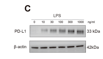
Application: WB
Reactivity: Human
Publish date: 2025 Mar
-
Citation
-
Nicotine-induced PD-L1 expression in lung squamous cell carcinoma is mediated by the α7-nAChR/STAT3 signaling pathway
Author: Yinan Li, Wenting Wang, Yaoyao Wang, Ce Zhou, Xin Zou, Yixuan Wang, Ning Wang
PMID: 10.21037/tcr-2024-2587
Journal: Translational Cancer Research
Application: WB
Reactivity: Human
Publish date: 2025 Jun
-
Citation
-
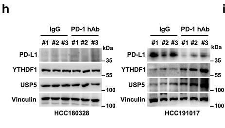
USP5 stabilizes YTHDF1 to control cancer immune surveillance through mTORC1-mediated phosphorylation
Author: Shao Na, Xi Lei, Lv Yangfan, Idris Muhammad, Zhang Lin, Cao Ya, Xiang Jingyi, Xu Xi, Ong Belinda X., Zhang Qiongyi, Peng Xu, Yue Xiaoyan, Xu Feng, Liu Chungang
PMID: 39900921

Journal: Nature Communications
Application: WB
Reactivity: Human,Mouse
Publish date: 2025 Feb
-
Citation
-
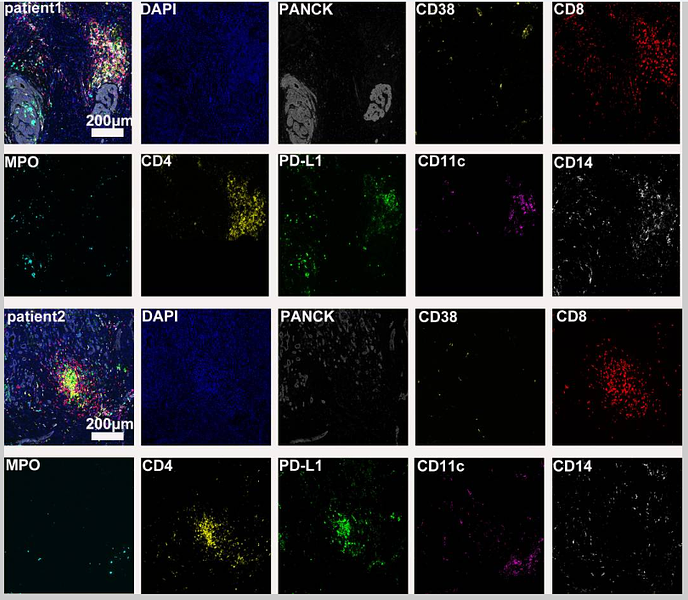
Case report: Diverse immune responses in advanced pancreatic ductal adenocarcinoma treated with immune checkpoint inhibitor-based conversion therapies
Author: Li Xiaoying, Xiao Chaoxin, Li Ruizhen, Zhang Pei, Cao Dan
PMID: 38415262

Journal: Frontiers In Immunology
Application: mIHC
Reactivity: Human
Publish date: 2024 Feb
-
Citation
Products with the same target and pathway
PD-L1 Recombinant Rabbit Monoclonal Antibody [PD00-97] - BSA and Azide free
Application: IHC-P,IF-Cell
Reactivity: Human
Conjugate: unconjugated
Human PD-L1 Recombinant Rabbit Monoclonal Antibody [PSH15-36] - BSA and Azide free (Capture)
Application: ELISA(Cap)
Reactivity: Human,Monkey
Conjugate: unconjugated
PD-L1 Recombinant Rabbit Multiclonal Antibody [PSH13-23]
Application: WB
Reactivity: Human,Mouse
Conjugate: unconjugated
PD-L1 Recombinant Rabbit Monoclonal Antibody [JJ08-95] - BSA and Azide free
Application: WB,IHC-P
Reactivity: Human
Conjugate: unconjugated
PD-L1 Recombinant Rabbit Monoclonal Antibody [JJ08-95]
Application: WB,IHC-P
Reactivity: Human
Conjugate: unconjugated
PD-L1 Recombinant Rabbit Monoclonal Antibody [PD00-97]
Application: IHC-P,IF-Cell
Reactivity: Human
Conjugate: unconjugated
PD-L1 Recombinant Rabbit Monoclonal Antibody
Application: mIHC
Reactivity: Human
Conjugate: unconjugated
PD-L1 Recombinant Rabbit Multiclonal Antibody [PSH13-23] - BSA and Azide free
Application: WB
Reactivity: Human,Mouse
Conjugate: unconjugated
Human PD-L1 Recombinant Rabbit Monoclonal Antibody [PSH15-37] - BSA and Azide free (Detector)
Application: ELISA(Det)
Reactivity: Human,Monkey
Conjugate: unconjugated
Biotin Conjugated Human PD-L1 Recombinant Rabbit Monoclonal Antibody [PSH15-37] - Detector
Application: ELISA(Det),ELISA
Reactivity: Human,Monkey
Conjugate: Biotin


