F4/80 Recombinant Rabbit Monoclonal Antibody [PSH0-85]
Specification
Overview
Product Name
F4/80 Recombinant Rabbit Monoclonal Antibody [PSH0-85]
Antibody Type
Recombinant Rabbit monoclonal Antibody
Immunogen
Recombinant protein within mouse F4/80 aa 1-650 / 931.
Species Reactivity
Mouse, Rat
Validated Applications
WB, IHC-P, IF-Tissue, IHC-Fr, mIHC
Molecular Weight
Predicted band size: 102 kDa
Positive Control
Mouse kidney tissue lysate, RAW264.7 cell lysate, Rat kidney tissue lysate, Rat spleen tissue lysate, mouse liver tissue, mouse spleen tissue, rat liver tissue, rat spleen tissue, mouse lung tissue, rat lung tissue.
Conjugation
unconjugated
Clone Number
PSH0-85
RRID
Product Features
Form
Liquid
Storage Instructions
Shipped at 4℃. Store at +4℃ short term (1-2 weeks). It is recommended to aliquot into single-use upon delivery. Store at -20℃ long term.
Storage Buffer
PBS (pH7.4), 0.1% BSA, 40% Glycerol. Preservative: 0.05% Sodium Azide.
Isotype
IgG
Purification Method
Protein A affinity purified.
Application Dilution
-
WB
-
1:1,000
-
IHC-P
-
1:200-1:1,000
-
IF-Tissue
-
1:500
-
IHC-Fr
-
1:1,000
-
mIHC
-
1:500
Applications in Publications
Species in Publications
Target
Function
EGF-like module-containing mucin-like hormone receptor-like 1 also known as F4/80 is a protein encoded by the ADGRE1 gene. EMR1 is a member of the adhesion GPCR family. Adhesion GPCRs are characterized by an extended extracellular region often possessing N-terminal protein modules that is linked to a TM7 region via a domain known as the GPCR-Autoproteolysis INducing (GAIN) domain. EMR1 expression in human is restricted to eosinophils and is a specific marker for these cells. The murine homolog of EMR1, F4/80, is a well-known and widely used marker of murine macrophage populations. The N-terminal fragment (NTF) of EMR1 contains 4-6 Epidermal Growth Factor-like (EGF-like) domains in human and 4-7 EGF-like domains in the mouse. Utilizing F4/80 knockout mice, Lin et al. showed that F4/80 is not necessary for the development of tissue macrophages but is required for the induction of efferent CD8+ regulatory T cells needed for peripheral tolerance.
Background References
1. Deng R et al. Periosteal CD68+ F4/80+ Macrophages Are Mechanosensitive for Cortical Bone Formation by Secretion and Activation of TGF-β1. Adv Sci (Weinh). 2022 Jan
2. Shin AE et al. F4/80+Ly6Chigh Macrophages Lead to Cell Plasticity and Cancer Initiation in Colitis. Gastroenterology. 2023 Apr
Subcellular Location
Cell membrane.
Synonyms
ADGRE1 antibody
Adhesion G protein coupled receptor E1 antibody
Adhesion G protein-coupled receptor E1 antibody
AGRE1_HUMAN antibody
Cell surface glycoprotein EMR1 antibody
Cell surface glycoprotein F4/80 antibody
DD7A5 7 antibody
Egf like module containing mucin like hormone receptor like 1 antibody
Egf like module containing mucin like hormone receptor like sequence 1 antibody
EGF like module receptor 1 antibody
ExpandADGRE1 antibody
Adhesion G protein coupled receptor E1 antibody
Adhesion G protein-coupled receptor E1 antibody
AGRE1_HUMAN antibody
Cell surface glycoprotein EMR1 antibody
Cell surface glycoprotein F4/80 antibody
DD7A5 7 antibody
Egf like module containing mucin like hormone receptor like 1 antibody
Egf like module containing mucin like hormone receptor like sequence 1 antibody
EGF like module receptor 1 antibody
EGF TM7 antibody
EGF-like module receptor 1 antibody
EGF-like module-containing mucin-like hormone receptor-like 1 antibody
EGFTM7 antibody
EMR 1 antibody
EMR1 antibody
EMR1 hormone receptor antibody
Gpf480 antibody
Ly71 antibody
Lymphocyte antigen 71 antibody
TM7LN3 antibody
CollapseImages
-

Western blot analysis of F4/80 on different lysates with Rabbit anti-F4/80 antibody (HA721520) at 1/1,000 dilution.
Lane 1: Mouse kidney tissue lysate (no heat) (40 µg/Lane)
Lane 2: Mouse kidney tissue lysate (40 µg/Lane)
Lane 3: RAW264.7 cell lysate (no heat) (20 µg/Lane)
Lane 4: Rat kidney tissue lysate (no heat) (40 µg/Lane)
Notice: no heat means the lysate is not boiled.
Predicted band size: 102 kDa
Observed band size: 160 kDa
Exposure time: 59 seconds; ECL: K1801;
4-20% SDS-PAGE gel.
Proteins were transferred to a PVDF membrane and blocked with 5% NFDM/TBST for 1 hour at room temperature. The primary antibody (HA721520) at 1/1,000 dilution was used in primary antibody dilution (K1803) at 4℃ overnight. Goat Anti-Rabbit IgG - HRP Secondary Antibody (HA1001) at 1/50,000 dilution was used for 1 hour at room temperature. -

☑ Relative expression (RE)
Western blot analysis of F4/80 on different lysates with Rabbit anti-F4/80 antibody (HA721520) at 1/1,000 dilution.
Lane 1: RAW264.7 cell lysate (no heat) (20 µg/Lane)
Lane 2: L929 cell lysate (no heat) (negative) (20 µg/Lane)
Lane 3: Rat spleen tissue lysate (70℃ heat) (40 µg/Lane)
Notice: no heat means the lysate is not boiled.
Predicted band size: 102 kDa
Observed band size: 160 kDa
Exposure time: 2 minutes;
4-20% SDS-PAGE gel.
Proteins were transferred to a PVDF membrane and blocked with 5% NFDM/TBST for 1 hour at room temperature. The primary antibody (HA721520) at 1/1,000 dilution was used in 5% NFDM/TBST at 4℃ overnight. Goat Anti-Rabbit IgG - HRP Secondary Antibody (HA1001) at 1:100,000 dilution was used for 1 hour at room temperature. -

-

-

Immunohistochemical analysis of paraffin-embedded mouse liver tissue with Rabbit anti-F4/80 antibody (HA721520) at 1/1,000 dilution.
The section was pre-treated using heat mediated antigen retrieval with Tris-EDTA buffer (pH 9.0) for 20 minutes. The tissues were blocked in 1% BSA for 20 minutes at room temperature, washed with ddH2O and PBS, and then probed with the primary antibody (HA721520) at 1/1,000 dilution for 1 hour at room temperature. The detection was performed using an HRP conjugated compact polymer system. DAB was used as the chromogen. Tissues were counterstained with hematoxylin and mounted with DPX. -

Immunohistochemical analysis of paraffin-embedded mouse spleen tissue with Rabbit anti-F4/80 antibody (HA721520) at 1/1,000 dilution.
The section was pre-treated using heat mediated antigen retrieval with Tris-EDTA buffer (pH 9.0) for 20 minutes. The tissues were blocked in 1% BSA for 20 minutes at room temperature, washed with ddH2O and PBS, and then probed with the primary antibody (HA721520) at 1/1,000 dilution for 1 hour at room temperature. The detection was performed using an HRP conjugated compact polymer system. DAB was used as the chromogen. Tissues were counterstained with hematoxylin and mounted with DPX. -

Immunohistochemical analysis of paraffin-embedded rat liver tissue with Rabbit anti-F4/80 antibody (HA721520) at 1/1,000 dilution.
The section was pre-treated using heat mediated antigen retrieval with Tris-EDTA buffer (pH 9.0) for 20 minutes. The tissues were blocked in 1% BSA for 20 minutes at room temperature, washed with ddH2O and PBS, and then probed with the primary antibody (HA721520) at 1/1,000 dilution for 1 hour at room temperature. The detection was performed using an HRP conjugated compact polymer system. DAB was used as the chromogen. Tissues were counterstained with hematoxylin and mounted with DPX. -

Immunohistochemical analysis of paraffin-embedded rat spleen tissue with Rabbit anti-F4/80 antibody (HA721520) at 1/1,000 dilution.
The section was pre-treated using heat mediated antigen retrieval with Tris-EDTA buffer (pH 9.0) for 20 minutes. The tissues were blocked in 1% BSA for 20 minutes at room temperature, washed with ddH2O and PBS, and then probed with the primary antibody (HA721520) at 1/1,000 dilution for 1 hour at room temperature. The detection was performed using an HRP conjugated compact polymer system. DAB was used as the chromogen. Tissues were counterstained with hematoxylin and mounted with DPX. -

Immunohistochemical analysis of paraffin-embedded mouse lung tissue with Rabbit anti-F4/80 antibody (HA721520) at 1/500 dilution.
The section was pre-treated using heat mediated antigen retrieval with Tris-EDTA buffer (pH 9.0) for 20 minutes. The tissues were blocked in 1% BSA for 20 minutes at room temperature, washed with ddH2O and PBS, and then probed with the primary antibody (HA721520) at 1/500 dilution for 1 hour at room temperature. The detection was performed using an HRP conjugated compact polymer system. DAB was used as the chromogen. Tissues were counterstained with hematoxylin and mounted with DPX. -

Immunohistochemical analysis of paraffin-embedded rat lung tissue with Rabbit anti-F4/80 antibody (HA721520) at 1/2,000 dilution.
The section was pre-treated using heat mediated antigen retrieval with Tris-EDTA buffer (pH 9.0) for 20 minutes. The tissues were blocked in 1% BSA for 20 minutes at room temperature, washed with ddH2O and PBS, and then probed with the primary antibody (HA721520) at 1/2,000 dilution for 1 hour at room temperature. The detection was performed using an HRP conjugated compact polymer system. DAB was used as the chromogen. Tissues were counterstained with hematoxylin and mounted with DPX. -

Application: IF-tissue
Species: Mouse
Site: Liver
Sample: Paraffin-embedded section
Antibody concentration: 1/500 -

Application: IF-tissue
Species: Rat
Site: Liver
Sample: Paraffin-embedded section
Antibody concentration: 1/500 -

Application: IF-tissue
Species: Mouse
Site: spleen
Sample: Paraffin-embedded section
Antibody concentration: 1/500 -

Application: IF-tissue
Species: Mouse
Site: small intestine
Sample: Paraffin-embedded section
Antibody concentration: 1/500 -

mIHC analysis of mouse spleen tissue (Formalin/PFA-fixed paraffin-embedded sections) with Rabbit anti-F4/80 antibody (HA721520) at 1/500 dilution. The immunostaining was performed with the IRISKitCmTSA Kit (900808). Heat mediated antigen retrieval with Tris-EDTA buffer (pH 9.0) for 30 mins at 95℃. DAPI (blue) was used as a nuclear counter stain. Image acquisition was performed with Olympus VS200 Slide Scanner.
Please note: All products are "FOR RESEARCH USE ONLY AND ARE NOT INTENDED FOR DIAGNOSTIC OR THERAPEUTIC USE"
Citation
-
Molybdenum nanodots reprogram inflammatory-driven osteolysis via bone immune remodeling
Author: Zi Fu, Wanting Hao, Xichun Qin, Han Wang, Dong Xie, Meng Zhang, Dalong Ni
PMID: 10.1016/j.biomaterials.2025.123871
Journal: Biomaterials
Application: mIHC
Reactivity: Mouse
Publish date: 2025 Nov
-
Citation
-
Activation of HTR2B Suppresses Osteosarcoma Progression through the STAT1-NLRP3 Inflammasome Pathway and Promotes OASL1+ Macrophage Production to Enhance Antitumor Immunity
Author: Zhen Huang, Jiazhuang Zhu, Jianping Hu, Xingkai Wang, Xiaolong Ma, Enjie Xu, Kunpeng Zhu, Chunlin Zhang
PMID: 40387572
Journal: Advanced Science
Application: mIF
Reactivity: Mouse
Publish date: 2025 May
-
Citation
-
Smilax china L. Rhizome Extract Enhances Anti-Tumor Immune Responses by Resetting M2-Like Macrophages and Tumor-Associated Macrophages to M1-Like via ERK1/2 Signaling
Author: Yingxue Guo, Xiaochen Lin, Penghao Wang, Yingying Wang, Mengyun Chen, Shuiyan Tang, Lu Jin, Weiye Mao, Xia Liu, Qiyang Shou, Huiying Fu
PMID: 40383248
Journal: Journal Of Ethnopharmacology
Application: IF
Reactivity: Mouse
Publish date: 2025 May
-
Citation
-
Chiral Gold Nanoparticles Manipulate Osteoimmune Microenvironment via Macrophage Autophagy for Bone Regeneration
Author:
PMID: 40786657
Journal: Materials Today Bio
Application: IF
Reactivity: Mouse
Publish date: 2025 Jul
-
Citation
-
L-arginine modified mesoporous bioactive glass with ROS scavenging and NO release for periodontitis treatment
Author: Haiyan Yao, Emine Sumeyra Turali Emre, Yuan Fan, Jiaolong Wang, Feng Liu, Junchao Wei
PMID: 40046013
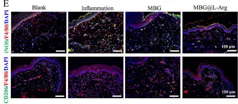
Journal: Bioactive Materials
Application: IF
Reactivity: Rat
Publish date: 2025 Feb
-
Citation
-
The extracellular-matrix-remodeling capability exposes therapeutic vulnerability in a subset of renal cell carcinomas with tumor thrombi
Author: Qi Zhang, Yezhen Tan, Qiyang Liang, Liangyou Gu, Yue Shi, Yan Huang, Xiubin Li, Jie Qi, Cheng Peng, Hanfeng Wang, Yaohui Wang, Kan Liu, Tianwei Cai, Yudan He, Qingbo Huang, Xu Zhang, Baojun Wang, Xin Ma, Weimin Ci
PMID: 40774251
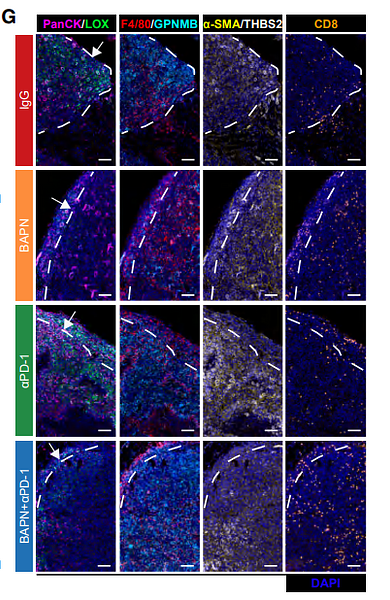
Journal: Developmental Cell
Application: mIHC
Reactivity: Mouse
Publish date: 2025 Aug
-
Citation
-
The insular cortex-nucleus tractus solitarius glutamatergic pathway involved in acute stress-induced gastric mucosal damage in rats
Author: Zepeng Wang, Xinyu Li, Yuanyuan Li, Xuehan Sun, Yuxue Wang, Tong Lu, Dongqin Zhao, Xiaoli Ma, Haiji Sun
PMID: 40242326
Journal: Neurobiology of Stress
Application: mIHC
Reactivity: Mouse
Publish date: 2025 Apr
-
Citation
-
Effect of macrophage-to-myofibroblast transition on silicosis
Author: Geng Fei,et al
PMID: 38979656
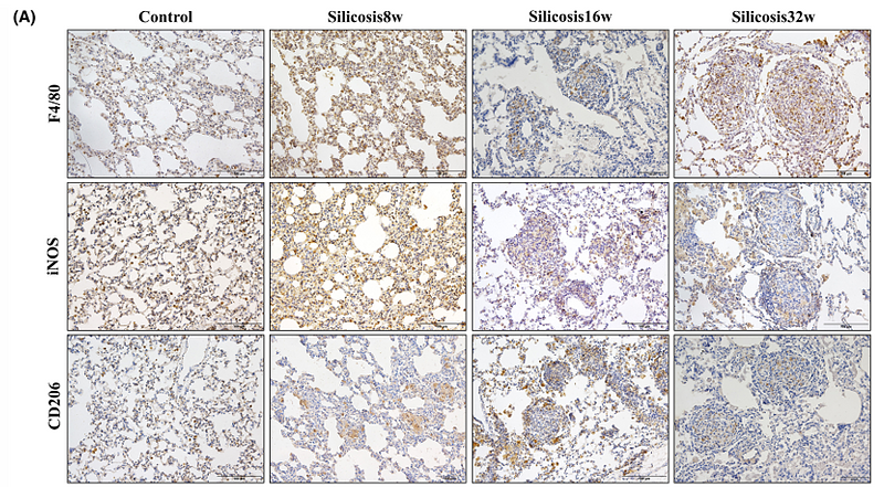
Journal: Animal Models And Experimental Medicine
Application: IHC-P
Reactivity: Mouse
Publish date: 2024 Jul
-
Citation
-
Calcium phosphate ceramic-induced osteoimmunomodulation: Submicron-surface-treated macrophage-derived exosomes driving osteogenesis
Author: Chen Fuying,et al
PMID: no pmid0501
Journal: Materials & Design
Application: IF
Reactivity: Mouse
Publish date: 2024 Apr
-
Citation
-
Integrated Bioinformatics Analysis and Experimental Verification of Immune Cell Infiltration and the Related Core Genes in Ulcerative Colitis
Author:
PMID: 37383675
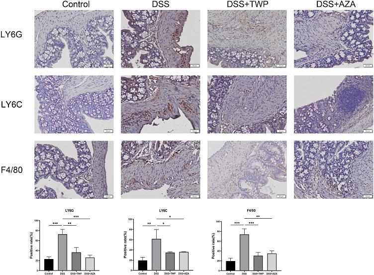
Journal: Pharmacogenomics & Personalized Medicine
Application: IHC-P
Reactivity: Mouse
Publish date: 2023 Jun
-
Citation
-
Ruthenium red attenuates acute pancreatitis by inhibiting MCU and improving mitochondrial function
Author:
PMID: 36283336

Journal: Biochemical And Biophysical Research Communications
Application: IHC-P
Reactivity: Mouse
Publish date: 2022 Dec
-
Citation
-
Complement C5 activation promotes type 2 diabetic kidney disease via activating STAT3 pathway and disrupting the gut‐kidney axis
Author:
PMID: 33280239
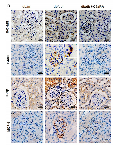
Journal: Journal Of Cellular And Molecular Medicine
Application: IHC-P
Reactivity: Mouse
Publish date: 2021 Jan
-
Citation

