AKT1/2/3 Recombinant Rabbit Monoclonal Antibody [JE75-09]
Catalog# HA721870
AKT1/2/3 Recombinant Rabbit Monoclonal Antibody [JE75-09]
-
WB
-
IF-Cell
-
FC
-
IP
-
Human
-
Mouse
-
Rat
-
Monkey
Overview
Product Name
AKT1/2/3 Recombinant Rabbit Monoclonal Antibody [JE75-09]
Antibody Type
Recombinant Rabbit monoclonal Antibody
Immunogen
Recombinant protein within Human AKT1/2/3 aa 231-480 / 480.
Species Reactivity
Human, Mouse, Rat, Monkey
Validated Applications
WB, IF-Cell, FC, IP
Molecular Weight
Predicted band size: 56 kDa
Positive Control
HeLa cell lysate, MCF7 cell lysate, A549 cell lysate, U-2 OS cell lysate, NIH/3T3 cell lysate, MCF7, COS-1 cell lysate, RAW264.7 cell lysate, C6 cell lysate, PC-12 cell lysate, Mouse brain tissue lysate, Mouse heart tissue lysate, Mouse testis tissue lysate, Rat brain tissue lysate, Rat heart tissue lysate, Rat testis tissue lysate, RAW264.7, C6.
Conjugation
unconjugated
Clone Number
JE75-09
RRID
Product Features
Form
Liquid
Concentration
1ug/ul
Storage Instructions
Store at +4℃ after thawing. Aliquot store at -20℃. Avoid repeated freeze / thaw cycles.
Storage Buffer
PBS (pH7.4), 0.1% BSA, 40% Glycerol. Preservative: 0.05% Sodium Azide.
Isotype
IgG
Purification Method
Protein A affinity purified.
Application Dilution
-
WB
-
1:2,000
-
IF-Cell
-
1:100
-
FC
-
1:1,000
-
IP
-
1-2μg/sample
Applications in Publications
Species in Publications
Target
Function
Akt, also referred to as PKB or Rac, plays a critical role in controlling cell survival and apoptosis. This protein kinase is activated by insulin and various growth and survival factors to function in a wortmannin-sensitive pathway involving PI3 kinase. Akt is activated by phospholipid binding and activation loop phosphorylation at Thr308 by PDK1 and by phosphorylation within the carboxy terminus at Ser473. The previously elusive PDK2 responsible for phosphorylation of Akt at Ser473 has been identified as mammalian target of rapamycin (mTOR) in a rapamycin-insensitive complex with rictor and Sin1 . Akt promotes cell survival by inhibiting apoptosis through phosphorylation and inactivation of several targets, including Bad, forkhead transcription factors, c-Raf, and caspase-9. PTEN phosphatase is a major negative regulator of the PI3K/Akt signaling pathway. LY294002 is a specific PI3 kinase inhibitor. Another essential Akt function is the regulation of glycogen synthesis through phosphorylation and inactivation of GSK-3α and β. Akt may also play a role in insulin stimulation of glucose transport. In addition to its role in survival and glycogen synthesis, Akt is involved in cell cycle regulation by preventing GSK-3β-mediated phosphorylation and degradation of cyclin D1 and by negatively regulating the cyclin-dependent kinase inhibitors p27 Kip1 and p21 Waf1/Cip1. Akt also plays a critical role in cell growth by directly phosphorylating mTOR in a rapamycin-sensitive complex containing raptor. More importantly, Akt phosphorylates and inactivates tuberin (TSC2), an inhibitor of mTOR within the mTOR-raptor complex.
Background References
1. Tian X et al. Costunolide is a dual inhibitor of MEK1 and AKT1/2 that overcomes osimertinib resistance in lung cancer. Mol Cancer. 2022 Oct
2. Chen Z et al. Nuclear DEK preserves hematopoietic stem cells potential via NCoR1/HDAC3-Akt1/2-mTOR axis. J Exp Med. 2021 May
Subcellular Location
Cytoplasm, Nucleus, Cell membrane.
UNIPROT
Synonyms
AKT antibody
AKT1 antibody
AKT1 kinase antibody
AKT1m antibody
AKT2 antibody
AKT2 kinase antibody
Akt3 antibody
AKT3_HUMAN antibody
CAKT antibody
CWS6 antibody
ExpandAKT antibody
AKT1 antibody
AKT1 kinase antibody
AKT1m antibody
AKT2 antibody
AKT2 kinase antibody
Akt3 antibody
AKT3_HUMAN antibody
CAKT antibody
CWS6 antibody
DKFZp434N0250 antibody
HIHGHH antibody
kinase Akt1 antibody
MGC99656 antibody
MPPH antibody
Murine thymoma viral (v-akt) homolog 2 antibody
pan-akt antibody
PKB ALPHA antibody
PKB antibody
PKB beta antibody
PKB gamma antibody
PKB-GAMMA antibody
PKB/Akt antibody
PKBALPHA antibody
PKBB antibody
PKBBETA antibody
PKBG antibody
PKBGAMMA antibody
PRKBA antibody
PRKBB antibody
PRKBG antibody
Protein kinase Akt 2 antibody
Protein kinase Akt-2 antibody
Protein kinase Akt-3 antibody
Protein kinase B alpha antibody
Protein kinase B antibody
Protein kinase B beta antibody
Protein kinase B gamma antibody
Proto oncogene c Akt antibody
Proto-oncogene c-Akt antibody
RAC ALPHA antibody
RAC alpha serine/threonine protein kinase antibody
RAC antibody
RAC BETA antibody
RAC beta serine/threonine protein kinase antibody
RAC PK alpha antibody
RAC PK beta antibody
rac protein kinase alpha antibody
rac protein kinase beta antibody
RAC-ALPHA antibody
RAC-alpha serine/threonine-protein kinase antibody
RAC-beta serine/threonine-protein kinase antibody
RAC-gamma antibody
RAC-gamma serine/threonine-protein kinase antibody
RAC-PK-alpha antibody
RAC-PK-beta antibody
RAC-PK-gamma antibody
RACALPHA antibody
RACalpha serine/threonine kinase antibody
RACBETA antibody
RACgamma antibody
RACgamma serine/threonine protein kinase antibody
RACPKgamma antibody
serine threonine protein kinase antibody
STK 2 antibody
STK-2 antibody
STK2 antibody
thymoma viral proto oncogene 1 antibody
thymoma viral proto oncogene antibody
V akt murine thymoma viral oncogene homolog 1 antibody
V akt murine thymoma viral oncogene homolog 2 antibody
V akt murine thymoma viral oncogene homolog 3 (protein kinase B, gamma) antibody
V akt murine thymoma viral oncogene homolog 3 antibody
V-AKT murine thymoma viral oncogene homolog 1 antibody
V-AKT murine thymoma viral oncogene homolog 2 antibody
V-AKT murine thymoma viral oncogene homolog 3 antibody
vakt murine thymoma viral oncogene homolog 1 antibody
vakt murine thymoma viral oncogene homolog 2 antibody
vakt murine thymoma viral oncogene homolog 3 antibody
CollapseImages
-

Western blot analysis of AKT1/2/3 on different lysates with Rabbit anti-AKT1/2/3 antibody (HA721870) at 1/2,000 dilution and competitor's antibody at 1/1,000 dilution.
Lane 1: HeLa cell lysate
Lane 2: MCF7 cell lysate
Lane 3: A549 cell lysate
Lane 4: U-2 OS cell lysate
Lane 5: NIH/3T3 cell lysate
Lysates/proteins at 15 µg/Lane.
Predicted band size: 56 kDa
Observed band size: 56 kDa
Exposure time: 30 seconds; ECL: K1802;
4-20% SDS-PAGE gel.
Proteins were transferred to a PVDF membrane and blocked with 5% NFDM/TBST for 1 hour at room temperature. The primary antibody (HA721870) at 1/2,000 dilution and competitor's antibody at 1/1,000 dilution were used in 5% NFDM/TBST at 4℃ overnight. Goat Anti-Rabbit IgG - HRP Secondary Antibody (HA1001) at 1/50,000 dilution was used for 1 hour at room temperature. -

Immunocytochemistry analysis of MCF7 cells labeling AKT1/2/3 with Rabbit anti-AKT1/2/3 antibody (HA721870) at 1/100 dilution.
Cells were fixed in 4% paraformaldehyde for 20 minutes at room temperature, permeabilized with 0.1% Triton X-100 in PBS for 5 minutes at room temperature, then blocked with 1% BSA in 10% negative goat serum for 1 hour at room temperature. Cells were then incubated with Rabbit anti-AKT1/2/3 antibody (HA721870) at 1/100 dilution in 1% BSA in PBST overnight at 4 ℃. Goat Anti-Rabbit IgG H&L (iFluor™ 488, HA1121) was used as the secondary antibody at 1/1,000 dilution. PBS instead of the primary antibody was used as the secondary antibody only control. Nuclear DNA was labelled in blue with DAPI.
Beta tubulin (M1305-2, red) was stained at 1/100 dilution overnight at +4℃. Goat Anti-Mouse IgG H&L (iFluor™ 594, HA1126) was used as the secondary antibody at 1/1,000 dilution. -

Western blot analysis of AKT1/2/3 on different lysates with Rabbit anti-AKT1/2/3 antibody (HA721870) at 1/2,000 dilution.
Lane 1: MCF7 cell lysate
Lane 2: A549 cell lysate
Lane 3: U-2 OS cell lysate
Lane 4: COS-1 cell lysate
Lane 5: NIH/3T3 cell lysate
Lane 6: RAW264.7 cell lysate
Lane 7: C6 cell lysate
Lane 8: PC-12 cell lysate
Lane 9: Mouse brain tissue lysate
Lane 10: Mouse heart tissue lysate
Lane 11: Mouse testis tissue lysate
Lane 12: Rat brain tissue lysate
Lane 13: Rat heart tissue lysate
Lane 14: Rat testis tissue lysate
Lysates/proteins at 20 µg/Lane.
Predicted band size: 56 kDa
Observed band size: 56 kDa
Exposure time: 2 minutes; ECL: K1801;
4-20% SDS-PAGE gel.
Proteins were transferred to a PVDF membrane and blocked with 5% NFDM/TBST for 1 hour at room temperature. The primary antibody (HA721870) at 1/2,000 dilution was used in 5% NFDM/TBST at room temperature for 2 hours. Goat Anti-Rabbit IgG - HRP Secondary Antibody (HA1001) at 1/50,000 dilution was used for 1 hour at room temperature. -

AKT1/2/3 was immunoprecipitated from 0.2 mg MCF7 cell lysate with HA721870 at 2 µg/25 µl agarose. Western blot was performed from the immunoprecipitate using HA721870 at 1/1,000 dilution. Anti-Rabbit IgG for IP Nano-secondary antibody (NBI01H) at 1/5,000 dilution was used for 1 hour at room temperature.
Lane 1: MCF7 cell lysate (input)
Lane 2: HA721870 IP in MCF7 cell lysate
Lane 3: Rabbit IgG instead of HA721870 in MCF7 cell lysate
Blocking/Dilution buffer: 5% NFDM/TBST
Exposure time: 6 seconds; ECL: K1801 -

Immunocytochemistry analysis of RAW264.7 cells labeling AKT1/2/3 with Rabbit anti-AKT1/2/3 antibody (HA721870) at 1/100 dilution.
Cells were fixed in 4% paraformaldehyde for 20 minutes at room temperature, permeabilized with 0.1% Triton X-100 in PBS for 5 minutes at room temperature, then blocked with 1% BSA in 10% negative goat serum for 1 hour at room temperature. Cells were then incubated with Rabbit anti-AKT1/2/3 antibody (HA721870) at 1/100 dilution in 1% BSA in PBST overnight at 4 ℃. Goat Anti-Rabbit IgG H&L (iFluor™ 488, HA1121) was used as the secondary antibody at 1/1,000 dilution. PBS instead of the primary antibody was used as the secondary antibody only control. Nuclear DNA was labelled in blue with DAPI.
Beta tubulin (M1305-2, red) was stained at 1/100 dilution overnight at +4℃. Goat Anti-Mouse IgG H&L (iFluor™ 594, HA1126) was used as the secondary antibody at 1/1,000 dilution. -

Immunocytochemistry analysis of C6 cells labeling AKT1/2/3 with Rabbit anti-AKT1/2/3 antibody (HA721870) at 1/100 dilution.
Cells were fixed in 4% paraformaldehyde for 20 minutes at room temperature, permeabilized with 0.1% Triton X-100 in PBS for 5 minutes at room temperature, then blocked with 1% BSA in 10% negative goat serum for 1 hour at room temperature. Cells were then incubated with Rabbit anti-AKT1/2/3 antibody (HA721870) at 1/100 dilution in 1% BSA in PBST overnight at 4 ℃. Goat Anti-Rabbit IgG H&L (iFluor™ 488, HA1121) was used as the secondary antibody at 1/1,000 dilution. PBS instead of the primary antibody was used as the secondary antibody only control. Nuclear DNA was labelled in blue with DAPI.
Beta tubulin (M1305-2, red) was stained at 1/100 dilution overnight at +4℃. Goat Anti-Mouse IgG H&L (iFluor™ 594, HA1126) was used as the secondary antibody at 1/1,000 dilution. -

Flow cytometric analysis of C6 cells labeling AKT1/2/3.
Cells were fixed and permeabilized. Then stained with the primary antibody (HA721870, 1/1,000) (red) compared with Rabbit IgG Isotype Control (green). After incubation of the primary antibody at +4℃ for an hour, the cells were stained with a iFluor™ 488 conjugate-Goat anti-Rabbit IgG Secondary antibody (HA1121) at 1/1,000 dilution for 30 minutes at +4℃. Unlabelled sample was used as a control (cells without incubation with primary antibody; black). -

Flow cytometric analysis of RAW264.7 cells labeling AKT1/2/3.
Cells were fixed and permeabilized. Then stained with the primary antibody (HA721870, 1/1,000) (red) compared with Rabbit IgG Isotype Control (green). After incubation of the primary antibody at +4℃ for an hour, the cells were stained with a iFluor™ 488 conjugate-Goat anti-Rabbit IgG Secondary antibody (HA1121) at 1/1,000 dilution for 30 minutes at +4℃. Unlabelled sample was used as a control (cells without incubation with primary antibody; black). -

☑ Knockdown (KD)
Western blot analysis of AKT1/2/3 on different lysates with Rabbit anti-AKT1/2/3 antibody (HA721870) at 1/2,000 dilution.
Lane 1: MCF7-si NT cell lysate
Lane 2: MCF7-si AKT1/2/3 cell lysate
Lysates/proteins at 10 µg/Lane.
Predicted band size: 56 kDa
Observed band size: 56 kDa
Exposure time: 7 seconds; ECL: K1801;
4-20% SDS-PAGE gel.
Proteins were transferred to a PVDF membrane and blocked with 5% NFDM/TBST for 1 hour at room temperature. The primary antibody (HA721870) at 1/2,000 dilution was used in 5% NFDM/TBST at 4℃ overnight. Goat Anti-Rabbit IgG - HRP Secondary Antibody (HA1001) at 1/50,000 dilution was used for 1 hour at room temperature.
Please note: All products are "FOR RESEARCH USE ONLY AND ARE NOT INTENDED FOR DIAGNOSTIC OR THERAPEUTIC USE"
Citation
-
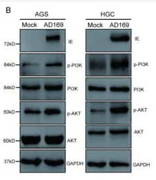
Human cytomegalovirus tegument protein UL23 promotes gastric cancer immune evasion by facilitating PD-L1 transcription
Author: Shiyu Feng, Yitian Shen, Haoke Zhang, Wanfeng Liu, Weixu Feng, Xiuting Chen, Liang Zhang, Jiangli Chen, Mingdong Lu, Xiangyang Xue, Xian Shen
PMID: 39934685

Journal: Molecular Medicine
Application: WB
Reactivity: Human
Publish date: 2025 Feb
-
Citation
-
Exploring the mechanism of Polygonum Cuspidatum in the treatment of ischemic stroke by network pharmacology analysis and experimental validation
Author: Xingqin Cao, Shiqing Zhang, Mingjiang Mao, Qianwen Zhang, Ying Guo
PMID: 39909363

Journal: Fitoterapia
Application: WB
Reactivity: Rat
Publish date: 2025 Feb
-
Citation
-
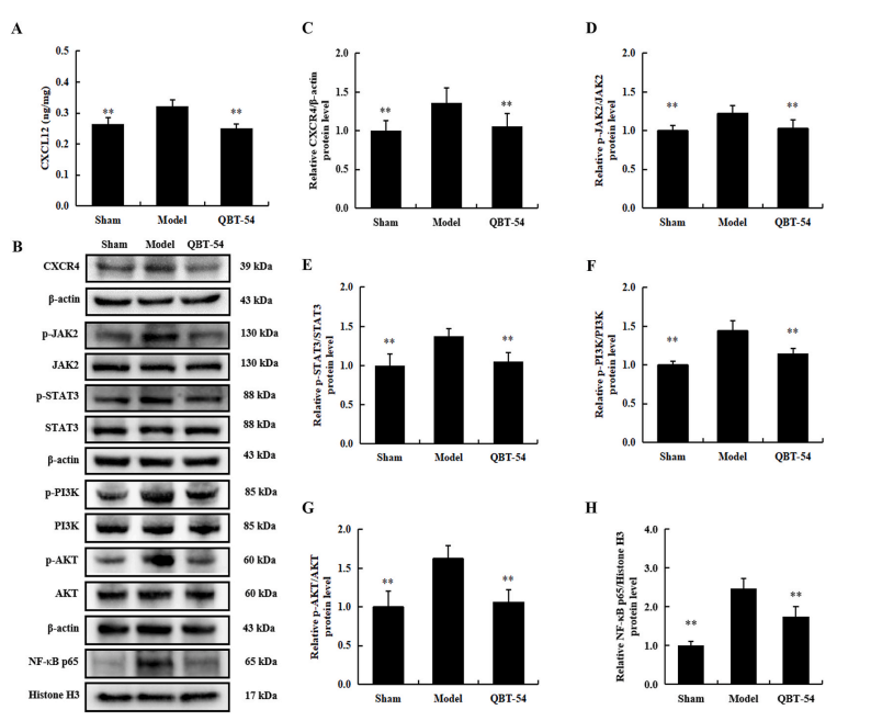
A novel phthalein component ameliorates neuroinflammation and cognitive dysfunction by suppressing the CXCL12/CXCR4 axis in rats with vascular dementia
Author: Kai-Ting Ma, Yi-Jin Wu, Yu-Xin Yang, Ting Wu, Chu Chen, Fu Peng, Jun-Rong Du, Cheng Peng
PMID: 38548120

Journal: Journal Of Ethnopharmacology
Application:
Reactivity:
Publish date: 2024 Mar
-
Citation
Products with the same target and pathway
AKT1/2/3 Recombinant Rabbit Monoclonal Antibody [ST48-09]
Application: WB,IF-Cell,IF-Tissue,IHC-P,IP,FC,IHC-Fr
Reactivity: Human,Mouse,Rat,Monkey
Conjugate: unconjugated
AKT1/2/3 Recombinant Rabbit Monoclonal Antibody [JE75-09] - BSA and Azide free
Application: WB,IF-Cell,FC,IP
Reactivity: Human,Mouse,Rat,Monkey
Conjugate: unconjugated
AKT1/2/3 Recombinant Rabbit Monoclonal Antibody [ST48-09] - BSA and Azide free
Application: WB,IF-Cell,IF-Tissue,IHC-P,IP,FC,IHC-Fr
Reactivity: Human,Mouse,Rat,Monkey
Conjugate: unconjugated
AKT1/2/3 Rabbit Polyclonal Antibody
Application: WB,IF-Cell,IHC-P,FC
Reactivity: Human,Mouse,Rat
Conjugate: unconjugated


