CD163 Recombinant Rabbit Monoclonal Antibody [JA51-30] - BSA and Azide free


Catalog# HA750412
CD163 Recombinant Rabbit Monoclonal Antibody [JA51-30] - BSA and Azide free
-
WB
-
IHC-P
-
IF-Tissue
-
IP
-
Human
-
ET1704-43
含抗保成分
-
unconjugated
Overview
Product Name
CD163 Recombinant Rabbit Monoclonal Antibody [JA51-30] - BSA and Azide free
Antibody Type
Recombinant Rabbit monoclonal Antibody
Immunogen
Recombinant protein within Human CD163 aa 1012-1149 / 1156.
Species Reactivity
Human
Validated Applications
WB, IHC-P, IF-Tissue, IP
Target Molecular Weight
Predicted band size: 125 kDa
Positive Control
Human pancreatic carcinoma, human cervical cancer, human lung tissue lysates, human liver tissue lysates, human liver tissue, human spleen tissue, human placenta tissue, human tonsil tissue.
Conjugation
unconjugated
Clone Number
JA51-30
Product Features
Form
Liquid
Concentration
Storage Instructions
Store at +4℃ after thawing. Aliquot store at -20℃ or -80℃. Avoid repeated freeze / thaw cycles.
Storage Buffer
1*PBS (pH7.4).
Isotype
IgG
Purification Method
Protein A affinity purified.
Application Dilution
-
WB
-
1:500-1:2,000
-
IHC-P
-
1:1,000-1:10,000
-
IF-Tissue
-
1:200
-
IP
-
Use at an assay dependent concentration.
Target
Function
CD163, also designated M130, is a macrophage-associated antigen that is a member of the scavenger receptor cysteine-rich (SRCR) superfamily. It is highly expressed on macrogphages and to a lesser extent on monocytes. The acute phase-regulated and signal-inducing macrophage protein, CD163, is a receptor that scavenges hemoglobin by mediating endocytosis of haptoglobin-hemoglobin complexes. CD163 binds only haptoglobin and hemoglobin in complex, which indicates the exposure of a receptor-binding neoepitope. The receptor-ligand interaction is calcium-dependent and of high affinity. The existence of several CD163 isoforms, which differ in the structure of their cytoplasmic domains and putative phosphorylation sites, suggests that these isoforms also differ in their signaling mechanism. The gene which encodes CD163 maps to human chromosome 12p13.31.
Background References
1. Rowland RRR et al. Role of CD163 in PRRSV infection. Virology. 2024 Dec
2. Mori M et al. CD163(+) Macrophages Induce Endothelial-to-Mesenchymal Transition in Atheroma. Circ Res. 2024 Jul
Tissue Specificity
Expressed in monocytes and mature macrophages such as Kupffer cells in the liver, red pulp macrophages in the spleen, cortical macrophages in the thymus, resident bone marrow macrophages and meningeal macrophages of the central nervous system. Expressed also in blood. Isoform 1 is the lowest abundant in the blood. Isoform 2 is the lowest abundant in the liver and the spleen. Isoform 3 is the predominant isoform detected in the blood.
Post-translational Modification
A soluble form (sCD163) is produced by proteolytic shedding which can be induced by lipopolysaccharide, phorbol ester and Fc region of immunoglobulin gamma. This cleavage is dependent on protein kinase C and tyrosine kinases and can be blocked by protease inhibitors. The shedding is inhibited by the tissue inhibitor of metalloproteinase TIMP3, and thus probably induced by membrane-bound metalloproteinases ADAMs.; Phosphorylated.
Subcellular Location
Cell membrane; Secreted.
UNIPROT
Synonyms
C163A_HUMAN antibody
CD 163 antibody
CD163 antibody
CD163 antigen antibody
CD163 molecule antibody
Hemoglobin scavenger receptor antibody
M130 antibody
M130 antigen precursor antibody
Macrophage associated antigen antibody
MM130 antibody
ExpandImages
-
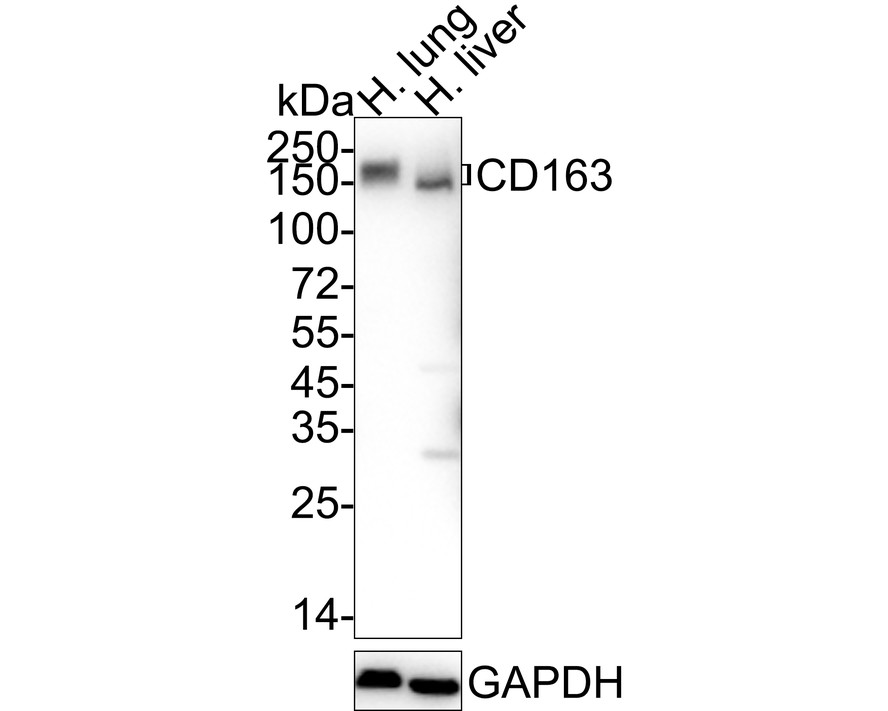
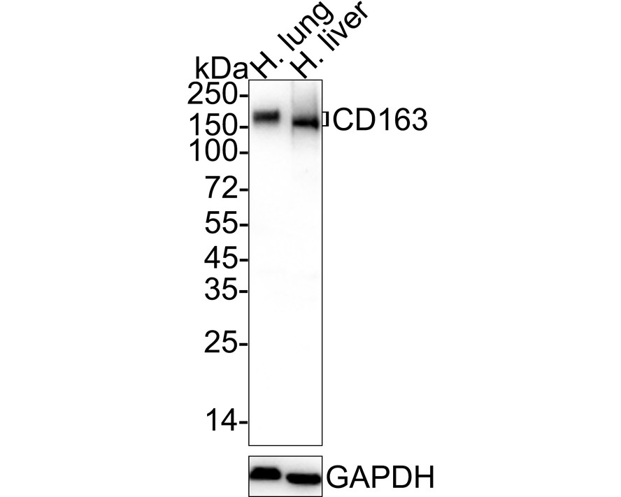
Western blot analysis of CD163 on different lysates with Rabbit anti-CD163 antibody (HA750412) at 1/1,000 dilution.
Lane 1: Human lung tissue lysate
Lane 2: Human liver tissue lysate
Lysates/proteins at 40 µg/Lane.
Predicted band size: 125 kDa
Observed band size: 150-170 kDa
Exposure time: 1 minute; ECL: K1801;
4-20% SDS-PAGE gel.
Proteins were transferred to a PVDF membrane and blocked with 5% NFDM/TBST for 1 hour at room temperature. The primary antibody (HA750412) at 1/1,000 dilution was used in 5% NFDM/TBST at 4℃ overnight. Goat Anti-Rabbit IgG - HRP Secondary Antibody (HA1001) at 1/50,000 dilution was used for 1 hour at room temperature. -
Immunohistochemical analysis of paraffin-embedded human liver tissue with Rabbit anti-CD163 antibody (HA750412) at 1/10,000 dilution.
The section was pre-treated using heat mediated antigen retrieval with Tris-EDTA buffer (pH 9.0) for 20 minutes. The tissues were blocked in 1% BSA for 20 minutes at room temperature, washed with ddH2O and PBS, and then probed with the primary antibody (HA750412) at 1/10,000 dilution for 1 hour at room temperature. The detection was performed using an HRP conjugated compact polymer system. DAB was used as the chromogen. Tissues were counterstained with hematoxylin and mounted with DPX. -
Immunohistochemical analysis of paraffin-embedded human spleen tissue with Rabbit anti-CD163 antibody (HA750412) at 1/10,000 dilution.
The section was pre-treated using heat mediated antigen retrieval with Tris-EDTA buffer (pH 9.0) for 20 minutes. The tissues were blocked in 1% BSA for 20 minutes at room temperature, washed with ddH2O and PBS, and then probed with the primary antibody (HA750412) at 1/10,000 dilution for 1 hour at room temperature. The detection was performed using an HRP conjugated compact polymer system. DAB was used as the chromogen. Tissues were counterstained with hematoxylin and mounted with DPX. -
Immunohistochemical analysis of paraffin-embedded human placenta tissue with Rabbit anti-CD163 antibody (HA750412) at 1/1,000 dilution.
The section was pre-treated using heat mediated antigen retrieval with Tris-EDTA buffer (pH 9.0) for 20 minutes. The tissues were blocked in 1% BSA for 20 minutes at room temperature, washed with ddH2O and PBS, and then probed with the primary antibody (HA750412) at 1/1,000 dilution for 1 hour at room temperature. The detection was performed using an HRP conjugated compact polymer system. DAB was used as the chromogen. Tissues were counterstained with hematoxylin and mounted with DPX. -
Immunohistochemical analysis of paraffin-embedded human tonsil tissue with Rabbit anti-CD163 antibody (HA750412) at 1/1,000 dilution.
The section was pre-treated using heat mediated antigen retrieval with Tris-EDTA buffer (pH 9.0) for 20 minutes. The tissues were blocked in 1% BSA for 20 minutes at room temperature, washed with ddH2O and PBS, and then probed with the primary antibody (HA750412) at 1/1,000 dilution for 1 hour at room temperature. The detection was performed using an HRP conjugated compact polymer system. DAB was used as the chromogen. Tissues were counterstained with hematoxylin and mounted with DPX. -
Application: IF-Tissue
Species: Human
Site: Spleen
Sample: Paraffin-embedded section
Antibody concentration: 1/200
Please note: All products are "FOR RESEARCH USE ONLY AND ARE NOT INTENDED FOR DIAGNOSTIC OR THERAPEUTIC USE"
Products with the same target and pathway

CD163 Mouse Monoclonal Antibody [A3B5]
Application: IHC-P,FC
Reactivity: Human
Conjugate: unconjugated

Human CD163 Recombinant Rabbit Monoclonal Antibody [PSH13-79] - BSA and Azide free (Detector)
Application: ELISA(Det)
Reactivity: Human
Conjugate: unconjugated

CD163 Recombinant Antibody - Rat IgG1 (Chimeric)
Application: mIHC
Reactivity: Human
Conjugate: unconjugated

CD163 Recombinant Antibody [JA51-30] - Rat IgG1 (Chimeric)
Application: IHC-P,mIHC
Reactivity: Human
Conjugate: unconjugated

CD163 Recombinant Rabbit Monoclonal Antibody [PSH18-98] - BSA and Azide free
Application: WB,IF-Cell,IHC-P
Reactivity: Human,Mouse,Rat
Conjugate: unconjugated
CD163 Mouse Monoclonal Antibody [A3B3]
Application: WB,IHC-P,FC
Reactivity: Human
Conjugate: unconjugated

Biotin Conjugated Human CD163 Recombinant Rabbit Monoclonal Antibody [PSH13-79] - Detector
Application: ELISA(Det),ELISA
Reactivity: Human
Conjugate: Biotin

CD163 Recombinant Rabbit Monoclonal Antibody [PSH18-98]
Application: WB,IF-Cell,IHC-P
Reactivity: Human,Mouse,Rat
Conjugate: unconjugated

Human CD163 Recombinant Rabbit Monoclonal Antibody [PSH13-80] - BSA and Azide free (Capture)
Application: ELISA(Cap)
Reactivity: Human
Conjugate: unconjugated

CD163 Recombinant Antibody [JA51-30] - Rat IgG1 (Chimeric) - BSA and Azide free
Application: IHC-P
Reactivity: Human
Conjugate: unconjugated

CD163 Recombinant Rabbit Monoclonal Antibody [JA51-30]
Application: WB,IHC-P,mIHC,IF-Tissue,IP
Reactivity: Human
Conjugate: unconjugated

Human CD163 Recombinant Rabbit Monoclonal Antibody [PSH13-78] - BSA and Azide free (Capture)
Application: ELISA(Cap)
Reactivity: Human
Conjugate: unconjugated

CD163 Recombinant Rabbit Monoclonal Antibody [PSH15-06]
Application: IHC-P,WB
Reactivity: Human
Conjugate: unconjugated

CD163 Recombinant Rabbit Monoclonal Antibody
Application: mIHC
Reactivity: Human
Conjugate: unconjugated