CD163 Mouse Monoclonal Antibody [A3B3]


Catalog# EM1901-90
CD163 Mouse Monoclonal Antibody [A3B3]
-
WB
-
IHC-P
-
FC
-
Human
-
unconjugated
Overview
Product Name
CD163 Mouse Monoclonal Antibody [A3B3]
Antibody Type
Mouse Monoclonal Antibody
Immunogen
Recombinant protein within human CD163 aa 1-170.
Species Reactivity
Human
Validated Applications
WB, IHC-P, FC
Target Molecular Weight
Predicted band size: 125 kDa
Positive Control
Human lung tissue lysate, human liver tissue lysate, human lung tissue, human liver carcinoma tissue, human colom tissue, human placenta tissue, HT-29.
Conjugation
unconjugated
Clone Number
A3B3
RRID
Product Features
Form
Liquid
Concentration
Storage Instructions
Shipped at 4℃. Store at +4℃ short term (1-2 weeks). It is recommended to aliquot into single-use upon delivery. Store at -20℃ long term.
Storage Buffer
1*PBS (pH7.4), 0.2% BSA, 50% Glycerol. Preservative: 0.05% Sodium Azide.
Isotype
IgG1
Purification Method
Protein G affinity purified.
Application Dilution
-
WB
-
1:500-1:2,000
-
IHC-P
-
1:100-1:500
-
FC
-
1:50-1:100
Target
Function
Acute phase-regulated receptor involved in clearance and endocytosis of hemoglobin/haptoglobin complexes by macrophages and may thereby protect tissues from free hemoglobin-mediated oxidative damage. Exhibits a higher affinity for complexes of hemoglobin and multimeric haptoglobin of HP*1F phenotype than for complexes of hemoglobin and dimeric haptoglobin of HP*1S phenotype. Induces a cascade of intracellular signals that involves tyrosine kinase-dependent calcium mobilization, inositol triphosphate production and secretion of IL6 and CSF1. After shedding, the soluble form (sCD163) may play an anti-inflammatory role, and may be a valuable diagnostic parameter for monitoring macrophage activation in inflammatory conditions. Intravenous lipopolysaccharide (LPS) produces a rapid rise of sCD163 in plasma of patient as it induces metalloproteinase-mediated shedding from monocytes surface. The soluble form (sCD163) in plasma is a novel parameter in diseases affecting macrophage function and monocyte/macrophage load in the body. The concentration of sCD163 is probably reflecting the number of macrophages of the 'alternative macrophage activation' phenotype with a high CD163 expression playing a major role in dampening the inflammatory response and scavenging components of damaged cells. This has initiated a number of clinical studies for evaluation of sCD163 as a disease marker in inflammatory conditions e.g. infection, autoimmune disease, transplantation, atherosclerosis and cancer.
Background References
1. Akila P. et. al. Retraction Notice to "CD163 AND ITS EXPANDING FUNCTIONAL REPERTOIRE". Clin Chim Acta. 2018 Dec.
2. Yang H. et. al. Identification of CD163 as an antiinflammatory receptor for HMGB1-haptoglobin complexes. JCI Insight. 2018 Dec.
Tissue Specificity
Expressed in monocytes and mature macrophages such as Kupffer cells in the liver, red pulp macrophages in the spleen, cortical macrophages in the thymus, resident bone marrow macrophages and meningeal macrophages of the central nervous system. Expressed also in blood. Isoform 1 is the lowest abundant in the blood. Isoform 2 is the lowest abundant in the liver and the spleen. Isoform 3 is the predominant isoform detected in the blood.
Post-translational Modification
A soluble form (sCD163) is produced by proteolytic shedding which can be induced by lipopolysaccharide, phorbol ester and Fc region of immunoglobulin gamma. This cleavage is dependent on protein kinase C and tyrosine kinases and can be blocked by protease inhibitors. The shedding is inhibited by the tissue inhibitor of metalloproteinase TIMP3, and thus probably induced by membrane-bound metalloproteinases ADAMs.; Phosphorylated.
Subcellular Location
Secreted, cell membrane.
UNIPROT
Synonyms
C163A_HUMAN antibody
CD 163 antibody
CD163 antibody
CD163 antigen antibody
CD163 molecule antibody
Hemoglobin scavenger receptor antibody
M130 antibody
M130 antigen precursor antibody
Macrophage associated antigen antibody
MM130 antibody
ExpandImages
-
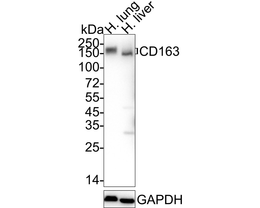
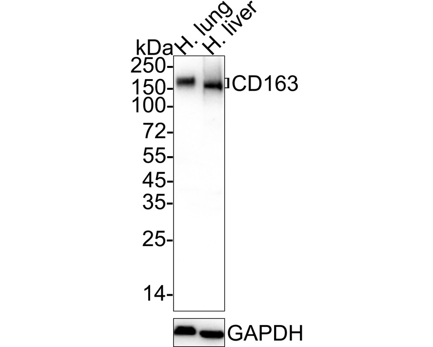
Western blot analysis of CD163 on different lysates with Mouse anti-CD163 antibody (EM1901-90) at 1/1,000 dilution.
Lane 1: Human lung tissue lysate
Lane 2: Human liver tissue lysate
Lysates/proteins at 40 µg/Lane.
Predicted band size: 125 kDa
Observed band size: 150-170 kDa
Exposure time: 25 seconds; ECL: K1801;
4-20% SDS-PAGE gel.
Proteins were transferred to a PVDF membrane and blocked with 5% NFDM/TBST for 1 hour at room temperature. The primary antibody (EM1901-90) at 1/1,000 dilution was used in 5% NFDM/TBST at 4℃ overnight. Goat Anti-Mouse IgG - HRP Secondary Antibody (HA1006) at 1/50,000 dilution was used for 1 hour at room temperature. -
Immunohistochemical analysis of paraffin-embedded human lung tissue using anti-CD163 antibody. The section was pre-treated using heat mediated antigen retrieval with Tris-EDTA buffer (pH 8.0-8.4) for 20 minutes.The tissues were blocked in 5% BSA for 30 minutes at room temperature, washed with ddH2O and PBS, and then probed with the primary antibody (EM1901-90, 1/200) for 30 minutes at room temperature. The detection was performed using an HRP conjugated compact polymer system. DAB was used as the chromogen. Tissues were counterstained with hematoxylin and mounted with DPX.
-
Immunohistochemical analysis of paraffin-embedded human liver carcinoma tissue using anti-CD163 antibody. The section was pre-treated using heat mediated antigen retrieval with Tris-EDTA buffer (pH 8.0-8.4) for 20 minutes.The tissues were blocked in 5% BSA for 30 minutes at room temperature, washed with ddH2O and PBS, and then probed with the primary antibody (EM1901-90, 1/200) for 30 minutes at room temperature. The detection was performed using an HRP conjugated compact polymer system. DAB was used as the chromogen. Tissues were counterstained with hematoxylin and mounted with DPX.
-
Immunohistochemical analysis of paraffin-embedded human colom tissue using anti-CD163 antibody. The section was pre-treated using heat mediated antigen retrieval with Tris-EDTA buffer (pH 8.0-8.4) for 20 minutes.The tissues were blocked in 5% BSA for 30 minutes at room temperature, washed with ddH2O and PBS, and then probed with the primary antibody (EM1901-90, 1/200) for 30 minutes at room temperature. The detection was performed using an HRP conjugated compact polymer system. DAB was used as the chromogen. Tissues were counterstained with hematoxylin and mounted with DPX.
-
Immunohistochemical analysis of paraffin-embedded human placenta tissue using anti-CD163 antibody. The section was pre-treated using heat mediated antigen retrieval with Tris-EDTA buffer (pH 8.0-8.4) for 20 minutes.The tissues were blocked in 5% BSA for 30 minutes at room temperature, washed with ddH2O and PBS, and then probed with the primary antibody (EM1901-90, 1/200) for 30 minutes at room temperature. The detection was performed using an HRP conjugated compact polymer system. DAB was used as the chromogen. Tissues were counterstained with hematoxylin and mounted with DPX.
-
Flow cytometric analysis of CD163 was done on HT-29 cells. The cells were fixed, permeabilized and stained with the primary antibody (EM1901-90, 1/50) (red). After incubation of the primary antibody at room temperature for an hour, the cells were stained with a Alexa Fluor 488-conjugated Goat anti-Mouse IgG Secondary antibody at 1/1000 dilution for 30 minutes.Unlabelled sample was used as a control (cells without incubation with primary antibody; black).
Please note: All products are "FOR RESEARCH USE ONLY AND ARE NOT INTENDED FOR DIAGNOSTIC OR THERAPEUTIC USE"
Citation
-
Squamous carcinoma cells drive lipid metabolic reprogramming of macrophages in head and neck squamous cell carcinoma by tunneling nanotube-mediated mitochondrial transfer
Journal: Journal of the National Cancer Center
DOI: 10.1016/j.jncc.2026.04.003
IF: 25.2
Application: WB
Reactivity: Human
Publish date: 2026 May
-
Crosstalk between cancer-associated fibroblasts and myeloid cells shape the heterogeneous microenvironment of gastric cancer
Journal: Current Genomics
DOI:
IF: 1.8
Application: IHC-P
Reactivity: Human
Publish date: 2024 Jul
-
Interactions between MFAP5 + fibroblasts and tumor-infiltrating myeloid cells shape the malignant microenvironment of colorectal cancer
Journal: Journal Of Translational Medicine
DOI:
IF: 8.440
Application: IHC-P
Reactivity: Human
Publish date: 2023 Jun
Products with the same target and pathway

CD163 Mouse Monoclonal Antibody [A3B5]
Application: IHC-P,FC
Reactivity: Human
Conjugate: unconjugated

Human CD163 Recombinant Rabbit Monoclonal Antibody [PSH13-79] - BSA and Azide free (Detector)
Application: ELISA(Det)
Reactivity: Human
Conjugate: unconjugated

Biotin Conjugated Human CD163 Recombinant Rabbit Monoclonal Antibody [PSH13-79] - Detector
Application: ELISA(Det),ELISA
Reactivity: Human
Conjugate: Biotin

Human CD163 Recombinant Rabbit Monoclonal Antibody [PSH13-80] - BSA and Azide free (Capture)
Application: ELISA(Cap)
Reactivity: Human
Conjugate: unconjugated

CD163 Recombinant Rabbit Monoclonal Antibody [JA51-30]
Application: WB,IHC-P,mIHC,IF-Tissue,IP
Reactivity: Human
Conjugate: unconjugated

CD163 Recombinant Rabbit Monoclonal Antibody [JA51-30] - BSA and Azide free
Application: WB,IHC-P,IF-Tissue,IP
Reactivity: Human
Conjugate: unconjugated

Human CD163 Recombinant Rabbit Monoclonal Antibody [PSH13-78] - BSA and Azide free (Capture)
Application: ELISA(Cap)
Reactivity: Human
Conjugate: unconjugated

CD163 Recombinant Rabbit Monoclonal Antibody [PSH15-06]
Application: IHC-P,WB
Reactivity: Human
Conjugate: unconjugated

CD163 Recombinant Rabbit Monoclonal Antibody
Application: mIHC
Reactivity: Human
Conjugate: unconjugated