Specification
Catalog# ET1601-23
GFAP Recombinant Rabbit Monoclonal Antibody [SA03-04]
-
WB
-
IHC-P
-
IF-Tissue
-
IHC-Fr
-
mIHC
-
IP
-
Human
-
Mouse
-
Rat
-
Cynomolgus monkey
-
Pig
-
unconjugated
Overview
Product Name
GFAP Recombinant Rabbit Monoclonal Antibody [SA03-04]
Antibody Type
Recombinant Rabbit monoclonal Antibody
Immunogen
Synthetic peptide within Human GFAP aa 1-50 / 432.
Species Reactivity
Human, Mouse, Rat (Predicted: Cynomolgus monkey, Pig)
Validated Applications
WB, IHC-P, IF-Tissue, IHC-Fr, mIHC, IP
Molecular Weight
Predicted band size: 50 kDa
Positive Control
Human brain tissue lysate, rat brain tissue lysate, human cerebellum tissue lysate, mouse brain tissue, rat brain tissue, human brain tissue, human glioblastoma tissue, mouse spinal cord tissue, rat spinal cord tissue, mouse cerebral cortex tissue, rat cerebral cortex tissue, mouse cerebellum tissue, rat cerebellum tissue, mouse hippocampus tissue, rat hippocampus tissue.
Conjugation
unconjugated
Clone Number
SA03-04
RRID
Product Features
Form
Liquid
Storage Instructions
Shipped at 4℃. Store at +4℃ short term (1-2 weeks). It is recommended to aliquot into single-use upon delivery. Store at -20℃ long term.
Storage Buffer
1*TBS (pH7.4), 0.05% BSA, 40% Glycerol. Preservative: 0.05% Sodium Azide.
Isotype
IgG
Purification Method
Protein A affinity purified.
Application Dilution
-
WB
-
1:5,000
-
IHC-P
-
1:1,000-1:2,000
-
IF-Tissue
-
1:500-1:1,000
-
IHC-Fr
-
1:500-1:1,000
-
mIHC
-
1:1,000-1:10,000
-
IP
-
Use at an assay dependent concentration.
Applications in Publications
| IF | See 12 publications below |
| WB | See 11 publications below |
| IF-tissue | See 3 publications below |
| IHC-P | See 1 publications below |
| IHC | See 1 publications below |
| IHC-Fr | See 1 publications below |
| IF-cell | See 1 publications below |
| IF-Tissue | See 1 publications below |
Species in Publications
Target
Function
Glial fibrillary acidic protein (GFAP) is a protein that is encoded by the GFAP gene in humans. It is a type III intermediate filament (IF) protein that is expressed by numerous cell types of the central nervous system (CNS), including astrocytes and ependymal cells during development. GFAP has also been found to be expressed in glomeruli and peritubular fibroblasts taken from rat kidneys, Leydig cells of the testis in both hamsters and humans, human keratinocytes, human osteocytes and chondrocytes and stellate cells of the pancreas and liver in rats. GFAP is closely related to the other three non-epithelial type III IF family members, vimentin, desmin and peripherin, which are all involved in the structure and function of the cell’s cytoskeleton. GFAP is thought to help to maintain astrocyte mechanical strength as well as the shape of cells, but its exact function remains poorly understood, despite the number of studies using it as a cell marker.
Background References
1. Zhang N et al. A self-assembly peptide nanofibrous scaffold reduces inflammatory response and promotes functional recovery in a mouse model of intracerebral hemorrhage. Nanomedicine N/A:N/A (2016).
2. Green AL et al. Preclinical antitumor efficacy of selective exportin 1 inhibitors in glioblastoma. Neuro Oncol 17:697-707 (2015).
Sequence Similarity
Belongs to the intermediate filament family.
Tissue Specificity
Expressed in cells lacking fibronectin.
Post-translational Modification
Phosphorylated by PKN1.
Subcellular Location
Cytoplasm
Synonyms
wu:fb34h11 antibody
ALXDRD antibody
cb345 antibody
etID36982.3 antibody
FLJ42474 antibody
FLJ45472 antibody
GFAP antibody
GFAP_HUMAN antibody
gfapl antibody
Glial fibrillary acidic protein antibody
Expandwu:fb34h11 antibody
ALXDRD antibody
cb345 antibody
etID36982.3 antibody
FLJ42474 antibody
FLJ45472 antibody
GFAP antibody
GFAP_HUMAN antibody
gfapl antibody
Glial fibrillary acidic protein antibody
Intermediate filament protein antibody
wu:fk42c12 antibody
xx:af506734 antibody
zgc:110485 antibody
CollapseImages
-

Application: IHC-Fr
Species: Mouse
Site: Hippocampus
Sample: Frozen section
Antibody concentration: 1/500
Antigen retrieval: Not required -

Application: IHC-Fr
Species: Rat
Site: Cerebral cortex
Sample: Frozen section
Antibody concentration: 1/500
Antigen retrieval: Not required -

Application: IF-tissue
Species: Mouse
Site: Cerebellum
Sample: Paraffin-embedded section
Antibody concentration: 1/500 -

Application: IF-tissue
Species: Rat
Site: Cerebral cortex
Sample: Paraffin-embedded section
Antibody concentration: 1/500 -

Fluorescence multiplex immunohistochemical analysis of mouse hippocampus (Formalin/PFA-fixed paraffin-embedded sections). Panel A: the merged image of anti-GFAP (ET1601-23, Green), anti-NeuN (ET1602-12, Red) and anti-c-Fos (HA722666, White) on hippocampus. HRP Conjugated UltraPolymer Goat Polyclonal Antibody HA1119/HA1120 was used as a secondary antibody. The immunostaining was performed with the Sequential Immuno-staining Kit (IRISKit™MH010101, www.luminiris.cn). The section was incubated in three rounds of staining: in the order of ET1601-23 (1/1,000 dilution), ET1602-12 (1/1,000 dilution) and HA722666 (1/200 dilution) for 20 mins at room temperature. Each round was followed by a separate fluorescent tyramide signal amplification system. Heat mediated antigen retrieval with Tris-EDTA buffer (pH 9.0) for 30 mins at 95℃. DAPI (blue) was used as a nuclear counter stain. Image acquisition was performed with Zeiss Observer 7 Inverted Fluorescence Microscope.
-

Fluorescence multiplex immunohistochemical analysis of mouse brain (Formalin/PFA-fixed paraffin-embedded sections). Panel A: the merged image of anti-GFAP (ET1601-23, Green), anti-NeuN (ET1602-12, Red) and anti-c-Fos (HA722666, White) on brain. HRP Conjugated UltraPolymer Goat Polyclonal Antibody HA1119/HA1120 was used as a secondary antibody. The immunostaining was performed with the Sequential Immuno-staining Kit (IRISKit™MH010101, www.luminiris.cn). The section was incubated in three rounds of staining: in the order of ET1601-23 (1/1,000 dilution), ET1602-12 (1/1,000 dilution) and HA722666 (1/200 dilution) for 20 mins at room temperature. Each round was followed by a separate fluorescent tyramide signal amplification system. Heat mediated antigen retrieval with Tris-EDTA buffer (pH 9.0) for 30 mins at 95℃. DAPI (blue) was used as a nuclear counter stain. Image acquisition was performed with Zeiss Observer 7 Inverted Fluorescence Microscope.
-

Fluorescence multiplex immunohistochemical analysis of mouse brain (Formalin/PFA-fixed paraffin-embedded sections). Panel A: the merged image of anti-MAP2 (HA500177, Red), anti-Olig2 (ET1604-29, Cyan), anti-GFAP (ET1601-23, Magenta) and anti-Neun (ET1602-12, Yellow) on mouse brain. HRP Conjugated UltraPolymer Goat Polyclonal Antibody HA1119/HA1120 was used as a secondary antibody. The immunostaining was performed with the Sequential Immuno-staining Kit (IRISKit™MH010101, www.luminiris.cn). The section was incubated in four rounds of staining: in the order of HA500177 (1/1,000 dilution), ET1604-29 (1/5,000 dilution), ET1601-23 (1/10,000 dilution) and ET1602-12 (1/10,000 dilution) for 20 mins at room temperature. Each round was followed by a separate fluorescent tyramide signal amplification system. Heat mediated antigen retrieval with Tris-EDTA buffer (pH 9.0) for 30 mins at 95℃. DAPI (blue) was used as a nuclear counter stain. Image acquisition was performed with Olympus VS200 Slide Scanner.
-

Fluorescence multiplex immunohistochemical analysis of mouse hippocampus (Formalin/PFA-fixed paraffin-embedded sections). Panel A: the merged image of anti-MAP2 (HA500177, Green), anti-GFAP (ET1601-23, Red) and anti-NeuN (ET1602-12, Magenta) on Mouse hippocampus. HRP Conjugated UltraPolymer Goat Polyclonal Antibody HA1119/HA1120 was used as a secondary antibody. The immunostaining was performed with the Sequential Immuno-staining Kit (IRISKit™MH010101, www.luminiris.cn). The section was incubated in three rounds of staining: in the order of HA500177 (1/1,000 dilution), ET1601-23 (1/1,000 dilution) and ET1602-12 (1/10,000 dilution) for 20 mins at room temperature. Each round was followed by a separate fluorescent tyramide signal amplification system. Heat mediated antigen retrieval with Tris-EDTA buffer (pH 9.0) for 30 mins at 95℃. DAPI (blue) was used as a nuclear counter stain. Image acquisition was performed with Olympus VS200 Slide Scanner.
-

Fluorescence multiplex immunohistochemical analysis of mouse brain (Formalin/PFA-fixed paraffin-embedded sections). Panel A: the merged image of anti-NeuN (ET1602-12, red), anti-Iba1 (ET1705-78, green), anti-GFAP (ET1601-23, gray), anti-Olig2 (ET1604-29, cyan), anti-MAP2 (HA500177, magenta) and anti-CD34 (ET1606-11, yellow) on mouse brain. HRP Conjugated UltraPolymer Goat Polyclonal Antibody HA1119/HA1120 was used as a secondary antibody. The immunostaining was performed with the Sequential Immuno-staining Kit (IRISKit™MH010101, www.luminiris.cn). The section was incubated in six rounds of staining: in the order of ET1602-12(1/5,000 dilution), ET1705-78 (1/2,000 dilution), ET1601-23 (1/5,000 dilution), ET1604-29 (1/1,000 dilution), HA500177 (1/5,000 dilution) and ET1606-11 (1/2,000 dilution) for 20 mins at room temperature. Each round was followed by a separate fluorescent tyramide signal amplification system. Heat mediated antigen retrieval with Tris-EDTA buffer (pH 9.0) for 30 mins at 95℃. DAPI (blue) was used as a nuclear counter stain. Image acquisition was performed with Olympus VS200 Slide Scanner.
-

Western blot analysis of GFAP on different lysates with Rabbit anti-GFAP antibody (ET1601-23) at 1/5,000 dilution.
Lane 1: Human brain tissue lysate
Lane 2: Rat brain tissue lysate
Lane 3: Human cerebellum tissue lysate
Lane 4: Mouse brain tissue lysate
Lysates/proteins at 20 µg/Lane.
Predicted band size: 50 kDa
Observed band size: 50 kDa
Exposure time: 11 seconds;
4-20% SDS-PAGE gel.
Proteins were transferred to a PVDF membrane and blocked with 5% NFDM/TBST for 1 hour at room temperature. The primary antibody (ET1601-23) at 1/5,000 dilution was used in 5% NFDM/TBST at 4℃ overnight. Goat Anti-Rabbit IgG - HRP Secondary Antibody (HA1001) at 1/50,000 dilution was used for 1 hour at room temperature. -

Immunohistochemical analysis of paraffin-embedded human brain tissue with Rabbit anti-GFAP antibody (ET1601-23) at 1/1,000 dilution.
The section was pre-treated using heat mediated antigen retrieval with Tris-EDTA buffer (pH 9.0) for 20 minutes. The tissues were blocked in 1% BSA for 20 minutes at room temperature, washed with ddH2O and PBS, and then probed with the primary antibody (ET1601-23) at 1/1,000 dilution for 1 hour at room temperature. The detection was performed using an HRP conjugated compact polymer system. DAB was used as the chromogen. Tissues were counterstained with hematoxylin and mounted with DPX. -

Immunohistochemical analysis of paraffin-embedded mouse cerebral cortex tissue with Rabbit anti-GFAP antibody (ET1601-23) at 1/1,000 dilution.
The section was pre-treated using heat mediated antigen retrieval with Tris-EDTA buffer (pH 9.0) for 20 minutes. The tissues were blocked in 1% BSA for 20 minutes at room temperature, washed with ddH2O and PBS, and then probed with the primary antibody (ET1601-23) at 1/1,000 dilution for 1 hour at room temperature. The detection was performed using an HRP conjugated compact polymer system. DAB was used as the chromogen. Tissues were counterstained with hematoxylin and mounted with DPX. -

Immunohistochemical analysis of paraffin-embedded rat cerebral cortex tissue with Rabbit anti-GFAP antibody (ET1601-23) at 1/1,000 dilution.
The section was pre-treated using heat mediated antigen retrieval with Tris-EDTA buffer (pH 9.0) for 20 minutes. The tissues were blocked in 1% BSA for 20 minutes at room temperature, washed with ddH2O and PBS, and then probed with the primary antibody (ET1601-23) at 1/1,000 dilution for 1 hour at room temperature. The detection was performed using an HRP conjugated compact polymer system. DAB was used as the chromogen. Tissues were counterstained with hematoxylin and mounted with DPX. -

Immunohistochemical analysis of paraffin-embedded rat cerebellum tissue with Rabbit anti-GFAP antibody (ET1601-23) at 1/1,000 dilution.
The section was pre-treated using heat mediated antigen retrieval with Tris-EDTA buffer (pH 9.0) for 20 minutes. The tissues were blocked in 1% BSA for 20 minutes at room temperature, washed with ddH2O and PBS, and then probed with the primary antibody (ET1601-23) at 1/1,000 dilution for 1 hour at room temperature. The detection was performed using an HRP conjugated compact polymer system. DAB was used as the chromogen. Tissues were counterstained with hematoxylin and mounted with DPX. -

Immunohistochemical analysis of paraffin-embedded mouse hippocampus tissue with Rabbit anti-GFAP antibody (ET1601-23) at 1/1,000 dilution.
The section was pre-treated using heat mediated antigen retrieval with Tris-EDTA buffer (pH 9.0) for 20 minutes. The tissues were blocked in 1% BSA for 20 minutes at room temperature, washed with ddH2O and PBS, and then probed with the primary antibody (ET1601-23) at 1/1,000 dilution for 1 hour at room temperature. The detection was performed using an HRP conjugated compact polymer system. DAB was used as the chromogen. Tissues were counterstained with hematoxylin and mounted with DPX. -

Immunohistochemical analysis of paraffin-embedded rat hippocampus tissue with Rabbit anti-GFAP antibody (ET1601-23) at 1/1,000 dilution.
The section was pre-treated using heat mediated antigen retrieval with Tris-EDTA buffer (pH 9.0) for 20 minutes. The tissues were blocked in 1% BSA for 20 minutes at room temperature, washed with ddH2O and PBS, and then probed with the primary antibody (ET1601-23) at 1/1,000 dilution for 1 hour at room temperature. The detection was performed using an HRP conjugated compact polymer system. DAB was used as the chromogen. Tissues were counterstained with hematoxylin and mounted with DPX. -

Immunohistochemical analysis of paraffin-embedded human brain tissue with Rabbit anti-GFAP antibody (ET1601-23) at 1/500 dilution.
Heat mediated antigen retrieval with Tris-EDTA buffer (pH 9.0, epitope retrieval solution 2) for 20 mins. The section was incubated with ET1601-23 for 30 mins at room temperature. The immunostaining was performed on a Leica Biosystems BOND® RX instrument. DAB was used as the chromogen. Tissues were counterstained with hematoxylin and mounted with DPX.
Please note: All products are "FOR RESEARCH USE ONLY AND ARE NOT INTENDED FOR DIAGNOSTIC OR THERAPEUTIC USE"
Citation
-
Integrin CD11b Alleviates Cerebral Ischemia/Reperfusion Injury via a Mechanism Involving Microglia/Macrophage Polarization
Author: Li Hui-Hua
PMID: 40974492
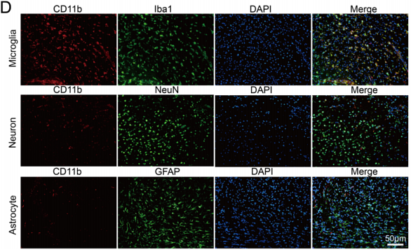
Journal: Journal of Molecular Neuroscience
Application: IF
Reactivity: Mouse
Publish date: 2025 Sept
-
Citation
-
Olfactory Ensheathing Cells Reduce Trigeminal Neuralgia Pain and Anxiety by Suppressing P2X7R-Mediated A1 Astrocyte Activation in Mice
Author: Jiafeng Lu, Zhonghua Chen, Xian Shao, Mengyun Li, Zhiwei Li, Lili Xu, Hui Lin, Lingyan He, Yue Shan
PMID: 41135808
Journal: Neuropharmacology
Application: IF-cell
Reactivity: Mouse
Publish date: 2025 Oct
-
Citation
-
G protein-coupled Estrogen Receptor Activation Exerts Protective Effects via Modulating Brain and Gut NLRP3 Inflammasome in Parkinson’s Disease
Author: Yan Liang, Liyuan Zhou, Hanqun Liu, Xiaoguang Huang, Yanhua Li, Xiaofeng Li, Shuxuan Huang
PMID: 41152022
Journal: Experimental Neurobiology
Application: IF-tissue,WB
Reactivity: Mouse
Publish date: 2025 Oct
-
Citation
-
5-Lipoxygenase triggers depressive-like behaviors via activating NLRP3-induced pyroptosis in CUMS-exposed mice
Author: Yani Zhang, Zheng Yang, Ting Xu, Qian Wang, Tingting Sang, Xuan Sheng, Yan Fang, Lisheng Chu
PMID: 41067520
Journal: Neuropharmacology
Application: IF-tissue
Reactivity: Mouse
Publish date: 2025 Oct
-
Citation
-
Icariin Supplementation Alleviates Cognitive Impairment Induced by d-Galactose via Modulation of the Gut–Brain Axis
Author: Siju Li, Menghui He, Ying He, Tingting Jin, Jianwen Chen, Junkang Peng, Wenhao Hu, Feng He
PMID: 40481799
Journal: Journal Of Agricultural And Food Chemistry
Application: IF
Reactivity: Mouse
Publish date: 2025 Jun
-
Citation
-
Fluorescence spectra of cell markers in the spinal cord for a murine model of amyotrophic lateral sclerosis with heat shock protein overexpression
Author: Gennadii A Piavchenko, Ksenia S Pokidova, Egor A Kuzmin, Artem A Venediktov, Igor Meglinski, Sergey L Kuznetsov
PMID: 21NOPMID25080101
Journal: Laser Physics Letters
Application: IF
Reactivity: Mouse
Publish date: 2025 Jun
-
Citation
-
Glioblastoma induces CAF-like astrocyte activation via the AKT/mTOR–SERPINH1/COL5A1 axis
Author: Jingxian Zhang, Yajia Chen, Hongwu Xu
PMID: 40530702
Journal: Biomolecules And Biomedicine
Application: IHC
Reactivity: Mouse
Publish date: 2025 Jun
-
Citation
-
Preventive and therapeutic effects of Tanshinone IIA on spinal cord injury without radiographic abnormality by regulating microglial phenotype polarization
Author: Luchun Xu, Yukun Ma, Guozheng Jiang, Zheng Cao, Jiawei Song, Yushan Gao, Guanlong Wang, Jiaojiao Fan, Yongdong Yang, Xing Yu
PMID: 40513327
Journal: International Immunopharmacology
Application: IF
Reactivity: Rat
Publish date: 2025 Jun
-
Citation
-
Neuronal TRPV1-CGRP axis regulates peripheral nerve regeneration through ERK/HIF-1 signaling pathway
Author: Huiling Che, Yu Du, Yixuan Jiang, Zhanfeng Zhu, Mingxuan Bai, Jianan Zheng, Mao Yang, Lin Xiang, Ping Gong
PMID: 39792906
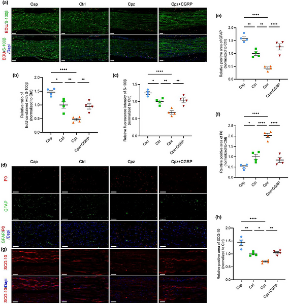
Journal: Journal Of Neurochemistry
Application: IF
Reactivity: Mouse
Publish date: 2025 Jan
-
Citation
-
Neuronal TRPV1-CGRP axis regulates peripheral nerve regeneration through ERK/HIF-1 signaling pathway
Author:
PMID: 10.1111/jnc.16281
Journal: JOURNAL OF NEUROCHEMISTRY
Application: IF-Tissue
Reactivity: Mouse
Publish date: 2025 Jan
-
Citation
-
Frequent fecal microbiota transplantation improves cognitive impairment and pathological changes in Alzheimer's disease FAD4T mice via the microbiota-gut-brain axis
Author: Zhiyan Zou, Dan Lei, Yuan Yin, Ruiling Xu, Huiwen Luo, Tingting Chen, Mingxue Liu, Xiaoan Li
PMID: 7NOPMID25040103
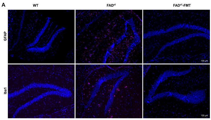
Journal: Heliyon
Application: IF,WB
Reactivity: Mouse
Publish date: 2025 Feb
-
Citation
-
Gastrodin Attenuates Cerebral Ischemia–Reperfusion Injury by Enhancing Mitochondrial Fusion and Activating the AMPK‐OPA1 Signaling Pathway
Author: Zihan Liu, Zeyu Han, Wenshuai Bao, Yihan Guo, Yuan Yuan, Jianming Cheng, Jie Zhang, Yang Hu
PMID: 10.1111/cns.70559
Journal: CNS Neuroscience & Therapeutics
Application: WB
Reactivity: Rat
Publish date: 2025 Aug
-
Citation
-
Poria cocos Polysaccharide Reshapes Gut Microbiota to Regulate Short-Chain Fatty Acids and Alleviate Neuroinflammation-Related Cognitive Impairment in Alzheimer’s Disease
Author: Meiying Song, Shanshan Zhang, Yuxin Gan, Tao Ding, Zhu Li, Xiang Fan
PMID: 40254847
Journal: Journal Of Agricultural And Food Chemistry
Application: IF
Reactivity: Mouse
Publish date: 2025 Apr
-
Citation
-
RNF13 protects neurons against ischemia-reperfusion injury via stabilizing p62-mediated Nrf2/HO-1 signaling pathway
Author: Qiangping Wang,et al
PMID: 39511649
Journal: Free Radical Biology And Medicine
Application: WB
Reactivity: Mouse
Publish date: 2024 Nov
-
Citation
-
Nitric oxide donor S‐Nitroso‐N‐acetyl penicillamine for hepatic stellate cells to restore quiescence
Author: Du Junbao,et al
PMID: NOPMID20240703
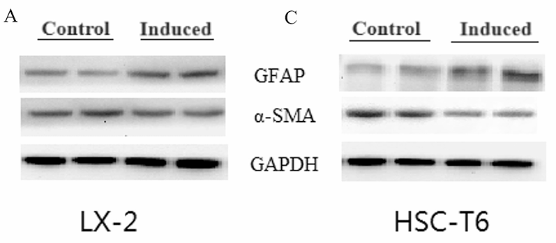
Journal: Pediatric Discovery
Application: IF,WB
Reactivity: Human,Rat
Publish date: 2024 Jul
-
Citation
-
Adenosine A2A receptor antagonist KW6002 protects against A53T mutant alpha-synuclein-induced brain damage and neuronal apoptosis in Parkinson's disease mice by restoring autophagic flux
Author: Hu Qidi,et al
PMID: 38157926
Journal: Neuroscience Letters
Application: IF-tissue
Reactivity: Mouse
Publish date: 2024 Jan
-
Citation
-
NKG2D knockdown improves hypoxic-ischemic brain damage by inhibiting neuroinflammation in neonatal mice
Author: Liu Lin,et al
PMID: 38282118
Journal: Scientific Reports
Application: IF
Reactivity: Mouse
Publish date: 2024 Feb
-
Citation
-
Identification and verification of key molecules in the epileptogenic process of focal cortical dysplasia
Author: Lingman Wang,et al
PMID: 39612062
Journal: Metabolic Brain Disease
Application: WB,IF
Reactivity: Rat
Publish date: 2024 Dec
-
Citation
-
Quantitative Immunofluorescence Mapping of HSP70’s Neuroprotective Effects in FUS-ALS Mouse Models
Author: Gennadii A. Piavchenko, Ksenia S. Pokidova, Egor A. Kuzmin, Artem A. Venediktov, Ilya Y. Izmailov, Igor V. Meglinski, Sergey L. Kuznetsov
PMID: 21NOPMID25020804
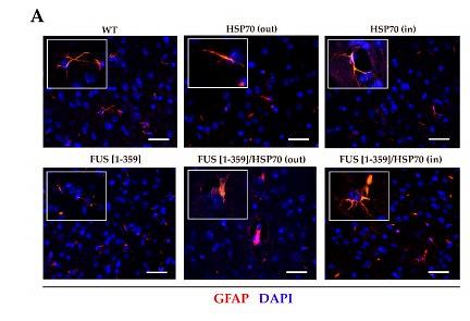
Journal: Applied Sciences-Basel
Application: IF
Reactivity: Mouse
Publish date: 2024 Dec
-
Citation
-
An oil-in-gel type of organohydrogel loaded with methylprednisolone for the treatment of secondary injuries following spinal cord traumas
Author: Yinqiu Tan, Ting Lai, Yuntao Li, Qi Tang, Weijia Zhang, Qi Liu, Sihan Wu, Xiao Peng, Xiaofeng Sui, Fulvio Reggiori, Xiaobing Jiang, Qianxue Chen, Cuifeng Wang
PMID: 39182693
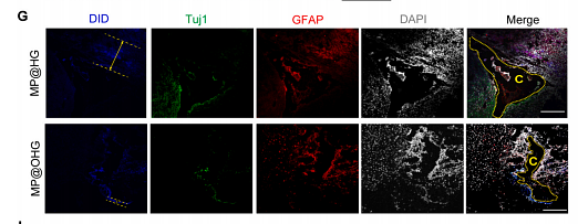
Journal: Journal Of Controlled Release
Application:
Reactivity:
Publish date: 2024 Aug
-
Citation
-
Small G-Protein Rheb Gates Mammalian Target of Rapamycin Signaling to Regulate Morphine Tolerance in Mice.
Author: Wang W, Ma X, Du W, Lin R, Li Z, Jiang W, Wang LY, Worley PF, Xu T
PMID: 38147625

Journal: Anesthesiology
Application: IHC-Fr
Reactivity: Mouse
Publish date: 2024 Apr
-
Citation
-
Deciphering the Therapeutic Potential of SheXiangXinTongNing: Interplay between Gut Microbiota and Brain Metabolomics in a CUMS Mice Model, with a Focus on Tryptophan Metabolism
Author: Wang Xiaohong,et al
PMID: 38704913
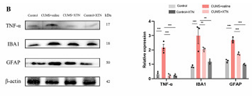
Journal: Phytomedicine
Application: WB
Reactivity: Mouse
Publish date: 2024 Apr
-
Citation
-
Catalpol rescues cognitive deficits by attenuating amyloid β plaques and neuroinflammation.
Author:
PMID: 37336148
Journal:
Application:
Reactivity: Mouse
Publish date: 2023 Sept
-
Citation
-
Astrocytic pyruvate dehydrogenase kinase-lactic acid axis involvement in glia-neuron crosstalk contributes to morphine-induced hyperalgesia in mice
Author: Xiaqing Ma, Qi Qi, Wenying Wang, Min Huang, Haiyan Wang, Limin Luo, Xiaotao Xu, Tifei Yuan, Haibo Shi, Wei Jiang, Tao Xu
PMID: 39161415
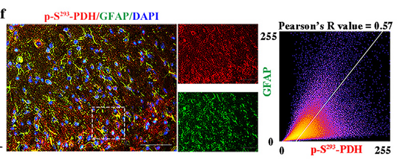
Journal: Fundamental Research
Application: WB
Reactivity: Mouse
Publish date: 2023 Mar
-
Citation
-
Modulation of microglial polarization by sequential targeting surface-engineered exosomes improves therapy for ischemic stroke
Author:
PMID: 37587291
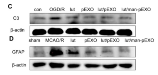
Journal: Drug Delivery And Translational Research
Application: WB
Reactivity: Human
Publish date: 2023 Aug
-
Citation
-
Multimodal investigation reveals the neuroprotective mechanism of Angong Niuhuang pill for intracerebral hemorrhage: Converging bioinformatics, network pharmacology,and experimental validation
Author:
PMID: 37633621
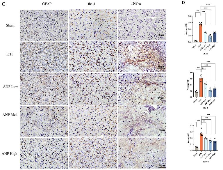
Journal: Journal Of Ethnopharmacology
Application: IHC-P,WB
Reactivity: Mouse
Publish date: 2023 Aug
-
Citation
-
The balance between AIM2-associated inflammation and autophagy: the role of CHMP2A in brain injury after cardiac arrest
Author: Shao, R., Wang, X., Xu, T., Xia, Y., & Cui, D.
PMID: 34740380
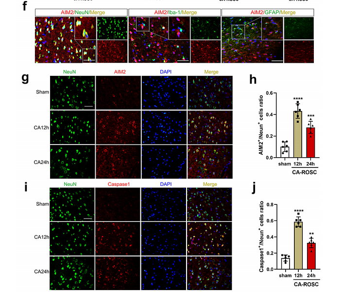
Journal: Journal Of Neuroinflammation
Application: IF
Reactivity: Rat
Publish date: 2021 Nov
-
Citation
-
Neuroprotective effects of saffron on the late cerebral ischemia injury through inhibiting astrogliosis and glial scar formation in rats
Author:
PMID: 32113053
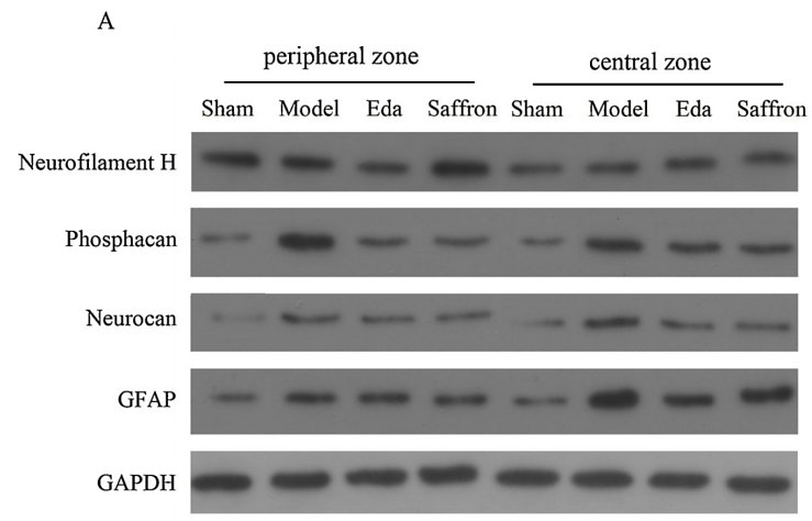
Journal: Biomedicine & Pharmacotherapy
Application: WB
Reactivity: Rat
Publish date: 2020 Jun
-
Citation
Products with the same target and pathway
iFluor™ 488 Conjugated GFAP Mouse Monoclonal Antibody [1-D4]
Application: IF-Tissue,IHC-Fr
Reactivity: Human,Rat
Conjugate: iFluor™ 488
GFAP Recombinant Mouse Monoclonal Antibody [PSH10-24] - BSA and Azide free
Application: WB,IHC-Fr,IHC-P,IF-Tissue
Reactivity: Human,Mouse,Rat
Conjugate: unconjugated
GFAP Mouse Monoclonal Antibody [1-D4]
Application: WB,IHC-P,IF-Tissue,IHC-Fr
Reactivity: Human,Rat
Conjugate: unconjugated
iFluor™ 594 Conjugated GFAP Mouse Monoclonal Antibody [1-D4]
Application: IF-Tissue
Reactivity: Human,Rat
Conjugate: iFluor™ 594
GFAP Recombinant Rabbit Monoclonal Antibody [PSH05-16] - BSA and Azide free (Capture)
Application: ELISA(Cap)
Reactivity: Human,Mouse,Rat
Conjugate: unconjugated
GFAP Recombinant Mouse Monoclonal Antibody [PSH05-17] - BSA and Azide free (Detector)
Application: ELISA(Det)
Reactivity: Human,Mouse,Rat
Conjugate: unconjugated
GFAP Rabbit Polyclonal Antibody
Application: WB,IF-Cell,IHC-P
Reactivity: Human,Mouse,Rat
Conjugate: unconjugated
Biotin Conjugated GFAP Mouse Monoclonal Antibody [PSH05-17]
Application: ELISA(Det)
Reactivity: Human,Mouse,Rat
Conjugate: Biotin
GFAP Rabbit Polyclonal Antibody
Application: WB,IF-Cell,IHC-P,FC
Reactivity: Human,Mouse,Rat
Conjugate: unconjugated
iFluor™ 647 Conjugated GFAP Mouse Monoclonal Antibody [1-D4]
Application: IF-Tissue
Reactivity: Human,Rat
Conjugate: iFluor™ 647
GFAP Recombinant Mouse Monoclonal Antibody [PSH10-24]
Application: WB,IHC-Fr,IHC-P,IF-Tissue
Reactivity: Human,Mouse,Rat
Conjugate: unconjugated


