MAP2 Rabbit Polyclonal Antibody
Specification
Catalog# HA500177
MAP2 Rabbit Polyclonal Antibody
-
WB
-
IF-Cell
-
IHC-P
-
mIHC
-
Human
-
Mouse
-
Rat
-
unconjugated
Overview
Product Name
MAP2 Rabbit Polyclonal Antibody
Antibody Type
Rabbit Polyclonal Antibody
Immunogen
Synthetic peptide within human MAP2 aa 1760-1810.
Species Reactivity
Human, Mouse, Rat
Validated Applications
WB, IF-Cell, IHC-P, mIHC
Molecular Weight
Predicted band size: 200 kDa
Positive Control
Mouse brain tissue lysates, rat brain tissue lysates, SH-SY5Y, human brain tissue, mouse brain tissue, rat brain tissue, human prostate carcinoma tissue, mouse hippocampus tissue.
Conjugation
unconjugated
RRID
Product Features
Form
Liquid
Storage Instructions
Shipped at 4℃. Store at +4℃ short term (1-2 weeks). It is recommended to aliquot into single-use upon delivery. Store at -20℃ long term.
Storage Buffer
1*TBS (pH7.4), 0.2% BSA, 50% Glycerol. Preservative: 0.05% Sodium Azide.
Isotype
IgG
Purification Method
Immunogen affinity purified.
Application Dilution
-
WB
-
1:1,000
-
IF-Cell
-
1:50-1:100
-
IHC-P
-
1:200-1:1,000
-
mIHC
-
1:1,000-1:5,000
Applications in Publications
| IF | See 5 publications below |
| WB | See 2 publications below |
| IF-cell | See 1 publications below |
| IF-Tissue | See 1 publications below |
Species in Publications
| Mouse | See 6 publications below |
| Rat | See 2 publications below |
| Human | See 2 publications below |
| Zebrafish | See 1 publications below |
Target
Function
This gene encodes a protein that belongs to the microtubule-associated protein family. The proteins of this family are thought to be involved in microtubule assembly, which is an essential step in neurogenesis. The products of similar genes in rat and mouse are neuron-specific cytoskeletal proteins that are enriched in dentrites, implicating a role in determining and stabilizing dentritic shape during neuron development. A number of alternatively spliced variants encoding distinct isoforms have been described.
Background References
1. DeGiosio R. et. al. MAP2 immunoreactivity deficit is conserved across the cerebral cortex within individuals with schizophrenia. NPJ Schizophr. 2019 Aug
2. Gumy LF. et. al. MAP2 Defines a Pre-axonal Filtering Zone to Regulate KIF1- versus KIF5-Dependent Cargo Transport in Sensory Neurons. Neuron. 2017 Apr
Subcellular Location
Cytoskeleton, dendrite.
Synonyms
DKFZp686I2148 antibody
MAP 2 antibody
MAP dendrite specific antibody
MAP-2 antibody
MAP2 antibody
MAP2A antibody
MAP2B antibody
MAP2C antibody
Microtubule associated protein 2 antibody
Microtubule-associated protein 2 antibody
ExpandDKFZp686I2148 antibody
MAP 2 antibody
MAP dendrite specific antibody
MAP-2 antibody
MAP2 antibody
MAP2A antibody
MAP2B antibody
MAP2C antibody
Microtubule associated protein 2 antibody
Microtubule-associated protein 2 antibody
MTAP2_HUMAN antibody
CollapseImages
-

Western blot analysis of MAP2 on different lysates with Rabbit anti-MAP2 antibody (HA500177) at 1/1,000 dilution.
Lane 1: Mouse brain tissue lysate
Lane 2: Rat brain tissue lysate
Lysates/proteins at 40 µg/Lane.
Predicted band size: 200 kDa
Observed band size: 300/70 kDa
Exposure time: 25 seconds; ECL: K1801;
4-20% SDS-PAGE gel.
Proteins were transferred to a PVDF membrane and blocked with 5% NFDM/TBST for 1 hour at room temperature. The primary antibody (HA500177) at 1/1,000 dilution was used in 5% NFDM/TBST at 4℃ overnight. Goat Anti-Rabbit IgG - HRP Secondary Antibody (HA1001) at 1/50,000 dilution was used for 1 hour at room temperature. -

Immunocytochemistry analysis of SH-SY5Y cells labeling MAP2 with Rabbit anti-MAP2 antibody (HA500177) at 1/50 dilution.
Cells were fixed in 4% paraformaldehyde for 10 minutes at 37 ℃, permeabilized with 0.05% Triton X-100 in PBS for 20 minutes, and then blocked with 2% negative goat serum for 30 minutes at room temperature. Cells were then incubated with Rabbit anti-MAP2 antibody (HA500177) at 1/50 dilution in 2% negative goat serum overnight at 4 ℃. Goat Anti-Rabbit IgG H&L (iFluor™ 488, HA1121) was used as the secondary antibody at 1/1,000 dilution. Nuclear DNA was labelled in blue with DAPI.
Beta tubulin (M1305-2, red) was stained at 1/200 dilution overnight at +4℃. Goat Anti-Mouse IgG H&L (iFluor™ 594, HA1126) was used as the secondary antibody at 1/1,000 dilution. -

Immunohistochemical analysis of paraffin-embedded human brain tissue with Rabbit anti-MAP2 antibody (HA500177) at 1/1,000 dilution.
The section was pre-treated using heat mediated antigen retrieval with Tris-EDTA buffer (pH 9.0) for 20 minutes. The tissues were blocked in 1% BSA for 20 minutes at room temperature, washed with ddH2O and PBS, and then probed with the primary antibody (HA500177) at 1/1,000 dilution for 1 hour at room temperature. The detection was performed using an HRP conjugated compact polymer system. DAB was used as the chromogen. Tissues were counterstained with hematoxylin and mounted with DPX. -

Immunohistochemical analysis of paraffin-embedded mouse brain tissue with Rabbit anti-MAP2 antibody (HA500177) at 1/1,000 dilution.
The section was pre-treated using heat mediated antigen retrieval with Tris-EDTA buffer (pH 9.0) for 20 minutes. The tissues were blocked in 1% BSA for 20 minutes at room temperature, washed with ddH2O and PBS, and then probed with the primary antibody (HA500177) at 1/1,000 dilution for 1 hour at room temperature. The detection was performed using an HRP conjugated compact polymer system. DAB was used as the chromogen. Tissues were counterstained with hematoxylin and mounted with DPX. -

Immunohistochemical analysis of paraffin-embedded rat brain tissue with Rabbit anti-MAP2 antibody (HA500177) at 1/1,000 dilution.
The section was pre-treated using heat mediated antigen retrieval with Tris-EDTA buffer (pH 9.0) for 20 minutes. The tissues were blocked in 1% BSA for 20 minutes at room temperature, washed with ddH2O and PBS, and then probed with the primary antibody (HA500177) at 1/1,000 dilution for 1 hour at room temperature. The detection was performed using an HRP conjugated compact polymer system. DAB was used as the chromogen. Tissues were counterstained with hematoxylin and mounted with DPX. -

Immunohistochemical analysis of paraffin-embedded human prostate carcinoma tissue using anti-MAP2 antibody. The section was pre-treated using heat mediated antigen retrieval with Tris-EDTA buffer (pH 8.0-8.4) for 20 minutes.The tissues were blocked in 5% BSA for 30 minutes at room temperature, washed with ddH2O and PBS, and then probed with the primary antibody (HA500177, 1/200) for 30 minutes at room temperature. The detection was performed using an HRP conjugated compact polymer system. DAB was used as the chromogen. Tissues were counterstained with hematoxylin and mounted with DPX.
-

Fluorescence multiplex immunohistochemical analysis of mouse brain (Formalin/PFA-fixed paraffin-embedded sections). Panel A: the merged image of anti-MAP2 (HA500177, Red), anti-Olig2 (ET1604-29, Cyan), anti-GFAP (ET1601-23, Magenta) and anti-Neun (ET1602-12, Yellow) on mouse brain. HRP Conjugated UltraPolymer Goat Polyclonal Antibody HA1119/HA1120 was used as a secondary antibody. The immunostaining was performed with the Sequential Immuno-staining Kit (IRISKit™MH010101, www.luminiris.cn). The section was incubated in four rounds of staining: in the order of HA500177 (1/1,000 dilution), ET1604-29 (1/5,000 dilution), ET1601-23 (1/10,000 dilution) and ET1602-12 (1/10,000 dilution) for 20 mins at room temperature. Each round was followed by a separate fluorescent tyramide signal amplification system. Heat mediated antigen retrieval with Tris-EDTA buffer (pH 9.0) for 30 mins at 95℃. DAPI (blue) was used as a nuclear counter stain. Image acquisition was performed with Olympus VS200 Slide Scanner.
-

Fluorescence multiplex immunohistochemical analysis of mouse hippocampus (Formalin/PFA-fixed paraffin-embedded sections). Panel A: the merged image of anti-MAP2 (HA500177, Green), anti-GFAP (ET1601-23, Red) and anti-NeuN (ET1602-12, Magenta) on Mouse hippocampus. HRP Conjugated UltraPolymer Goat Polyclonal Antibody HA1119/HA1120 was used as a secondary antibody. The immunostaining was performed with the Sequential Immuno-staining Kit (IRISKit™MH010101, www.luminiris.cn). The section was incubated in three rounds of staining: in the order of HA500177 (1/1,000 dilution), ET1601-23 (1/1,000 dilution) and ET1602-12 (1/10,000 dilution) for 20 mins at room temperature. Each round was followed by a separate fluorescent tyramide signal amplification system. Heat mediated antigen retrieval with Tris-EDTA buffer (pH 9.0) for 30 mins at 95℃. DAPI (blue) was used as a nuclear counter stain. Image acquisition was performed with Olympus VS200 Slide Scanner.
-

Fluorescence multiplex immunohistochemical analysis of mouse brain (Formalin/PFA-fixed paraffin-embedded sections). Panel A: the merged image of anti-NeuN (ET1602-12, red), anti-PAX6 (ET1612-58, green), anti-CD34 (ET1606-11, gray), anti-MAP2 (HA500177, magenta) and anti-TBR1 (ET1702-97, yellow) on mouse brain. HRP Conjugated UltraPolymer Goat Polyclonal Antibody HA1119/HA1120 was used as a secondary antibody. The immunostaining was performed with the Sequential Immuno-staining Kit (IRISKit™MH010101, www.luminiris.cn). The section was incubated in five rounds of staining: in the order of ET1602-12 (1/5,000 dilution), ET1612-58 (1/1,000 dilution), ET1606-11 (1/2,000 dilution), HA500177 (1/5,000 dilution) and ET1702-97 (1/1,000 dilution) for 20 mins at room temperature. Each round was followed by a separate fluorescent tyramide signal amplification system. Heat mediated antigen retrieval with Tris-EDTA buffer (pH 9.0) for 30 mins at 95℃. DAPI (blue) was used as a nuclear counter stain. Image acquisition was performed with Olympus VS200 Slide Scanner.
-

Fluorescence multiplex immunohistochemical analysis of mouse brain (Formalin/PFA-fixed paraffin-embedded sections). Panel A: the merged image of anti-NeuN (ET1602-12, red), anti-Iba1 (ET1705-78, green), anti-GFAP (ET1601-23, gray), anti-Olig2 (ET1604-29, cyan), anti-MAP2 (HA500177, magenta) and anti-CD34 (ET1606-11, yellow) on mouse brain. HRP Conjugated UltraPolymer Goat Polyclonal Antibody HA1119/HA1120 was used as a secondary antibody. The immunostaining was performed with the Sequential Immuno-staining Kit (IRISKit™MH010101, www.luminiris.cn). The section was incubated in six rounds of staining: in the order of ET1602-12(1/5,000 dilution), ET1705-78 (1/2,000 dilution), ET1601-23 (1/5,000 dilution), ET1604-29 (1/1,000 dilution), HA500177 (1/5,000 dilution) and ET1606-11 (1/2,000 dilution) for 20 mins at room temperature. Each round was followed by a separate fluorescent tyramide signal amplification system. Heat mediated antigen retrieval with Tris-EDTA buffer (pH 9.0) for 30 mins at 95℃. DAPI (blue) was used as a nuclear counter stain. Image acquisition was performed with Olympus VS200 Slide Scanner.
Please note: All products are "FOR RESEARCH USE ONLY AND ARE NOT INTENDED FOR DIAGNOSTIC OR THERAPEUTIC USE"
Citation
-
The role of REST in regulating the BDNF/TrkB signalling pathway in nano-alumina induced cognitive dysfunction in zebrafish
Author: Qiao Niu
PMID: 40912099
Journal: Ecotoxicology And Environmental Safety
Application: WB
Reactivity: Zebrafish
Publish date: 2025 Sept
-
Citation
-
A novel GSK3β inhibitor ameliorates tau aggregation and neuroinflammation in Alzheimer's disease
Author: Xin-Yue Ning, Wen-Jie Liu, Li-Jun Zhou, Nan Wang, Xin-Zhu Li, Li-Meng Wu, Zhen-Shu Li, Ai-Zhu Yang, Si-Yuan Liu, Zong-He Xu, Fang-Hua Xun, Zi-Hua Xu, Qing-Chun Zhao
PMID: 10.1016/j.neuint.2025.106090
Journal: Neurochemistry International
Application: IF-cell
Reactivity: Mouse
Publish date: 2025 Nov
-
Citation
-
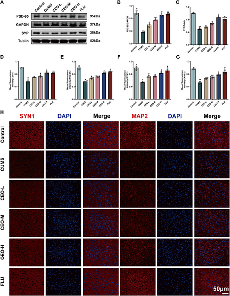
Rescue of CUMS-induced HPA axis hyperfunction and hypothalamic synaptic deficits by Citrus aurantium L. cv. Daidai essential oil via the cAMP/PKA/Grin2b pathway
Author:
PMID: 10.1016/j.jep.2025.119423
Journal: JOURNAL OF ETHNOPHARMACOLOGY
Application: IF-Tissue
Reactivity: Mouse
Publish date: 2025 Jan
-
Citation
-
Rescue of CUMS-induced HPA axis hyperfunction and hypothalamic synaptic deficits by Citrus aurantium L. cv. Daidai essential oil via the cAMP/PKA/Grin2b pathway
Author: Ze-Yu Zhang, Yu-Fei Liu, Si-Jia Zhang, Pan-Pan Zhang, Xiao-Xia Shen, Ji-Le Lan, Zhu-Jun Mao, Min-Jia Zhang, Ye-Ping Ruan, Xin Zhang
PMID: 39894418

Journal: Journal Of Ethnopharmacology
Application: IF
Reactivity: Mouse
Publish date: 2025 Jan
-
Citation
-
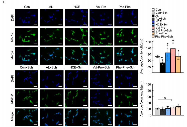
Hydrolyzed Chicken Meat Extract and Its Effective Cyclopeptides Promote Neurodevelopment by Activating Dopamine D1 Receptor and cAMP/PKA/CREB Pathway
Author: Xinyang Hu, Yifan Zheng, Haojie Jin, Yi Zhang, Wenlong Yang, Kunying Ding, Zhengwei Fu, Yinhua Ni
PMID: 8NOPMID25093001

Journal: Food Bioscience
Application: IF
Reactivity: Human,Mouse
Publish date: 2025 Aug
-
Citation
-
Mannose modified graphene oxide drug-delivery system targets cancer stem cells and tumor-associated macrophages to promote immunotherapeutic efficacy
Author: Jiapu Wang, Ziwei Liang, Yuhui Wang, Qi Liu, Shaojie Wang, Jie Wang, Ruxin Duan, Liqin Zhao, Yan Wei, Di Huang
PMID: 40286606
Journal: Colloids And Surfaces B: Biointerfaces
Application: IF
Reactivity: Human,Mouse
Publish date: 2025 Apr
-
Citation
-
Microenvironment-responsive injectable hydrogel for neuro-vascularized bone regeneration
Author: Wanshun Wang,et al
PMID: 39687796
Journal: Materials Today Bio
Application: IF
Reactivity: Rat
Publish date: 2024 Nov
-
Citation
-
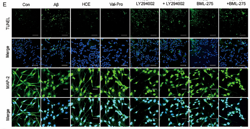
Hydrolyzed Chicken Meat Extract and Its Bioactive Cyclopeptides Protect Neural Function by Attenuating Inflammation and Apoptosis via PI3K/AKT and AMPK Pathways
Author: Ni Yinhua,et al
PMID: 39016108

Journal: Journal Of Agricultural And Food Chemistry
Application: IF
Reactivity: Mouse
Publish date: 2024 Jul
-
Citation
-
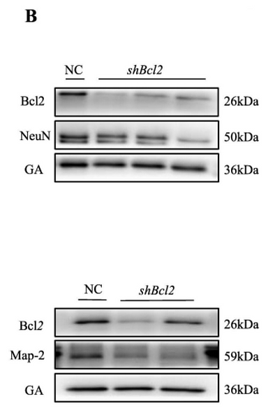
Potential role of Bcl2 in lipid metabolism and synaptic dysfunction of age-related hearing loss
Author: Yue Liu , Huasong Zhang , Cong Fan , Feiyi Liu , Shaoying Li , Juanjuan Li , Huiying Zhao , Xianhai Zeng
PMID: 37813166

Journal: Neurobiology Of Disease
Application: WB
Reactivity: Rat
Publish date: 2023 Oct
-
Citation
Alternative Products
MAP2 Recombinant Rabbit Monoclonal Antibody [PSH08-73]
Application: WB,IHC-P,IHC-Fr,IF-Cell,mIHC
Reactivity: Human,Mouse,Rat,Cynomolgus monkey
Conjugate: unconjugated
Products with the same target and pathway
MAP2 Recombinant Antibody [PSH08-73] - Rat IgG1 (Chimeric) - BSA and Azide free
Application: IHC-Fr,IHC-P,WB,IF-Cell
Reactivity: Human,Mouse,Rat
Conjugate: unconjugated
MAP2 Recombinant Antibody [PSH08-73] - Rat IgG1 (Chimeric)
Application: IHC-Fr,IHC-P,WB,IF-Cell
Reactivity: Human,Mouse,Rat
Conjugate: unconjugated
MAP2 Recombinant Rabbit Monoclonal Antibody [PSH08-73] - BSA and Azide free
Application: WB,IHC-P,IHC-Fr,IF-Cell
Reactivity: Human,Mouse,Rat,Cynomolgus monkey
Conjugate: unconjugated
MAP2 Recombinant Rabbit Monoclonal Antibody [PSH08-73]
Application: WB,IHC-P,IHC-Fr,IF-Cell,mIHC
Reactivity: Human,Mouse,Rat,Cynomolgus monkey
Conjugate: unconjugated


