FAK Recombinant Rabbit Monoclonal Antibody [SR46-04]

Specification
Catalog# ET1602-25
FAK Recombinant Rabbit Monoclonal Antibody [SR46-04]
-
WB
-
IF-Cell
-
IF-Tissue
-
IHC-P
-
FC
-
IP
-
Human
-
Mouse
-
Rat
Overview
Product Name
FAK Recombinant Rabbit Monoclonal Antibody [SR46-04]
Antibody Type
Recombinant Rabbit monoclonal Antibody
Immunogen
Synthetic peptide within human FAK aa 700-740.
Species Reactivity
Human, Mouse, Rat
Validated Applications
WB, IF-Cell, IF-Tissue, IHC-P, FC, IP
Molecular Weight
Predicted band size: 119 kDa
Positive Control
A431 cell lysate, U-87 MG cell lysate, NIH/3T3 cell lysate, C2C12 cell lysate, C6 cell lysate, C2C12, PANC-1, SH-SY5Y, human brain tissue, mouse brain tissue, rat brain tissue, rat kidney tissue, rat spleen tissue.
Conjugation
unconjugated
Clone Number
SR46-04
RRID
Product Features
Form
Liquid
Storage Instructions
Shipped at 4℃. Store at +4℃ short term (1-2 weeks). It is recommended to aliquot into single-use upon delivery. Store at -20℃ long term.
Storage Buffer
1*TBS (pH7.4), 0.05% BSA, 40% Glycerol. Preservative: 0.05% Sodium Azide.
Isotype
IgG
Purification Method
Protein A affinity purified.
Application Dilution
-
WB
-
1:5,000
-
IF-Cell
-
1:50-1:100
-
IF-Tissue
-
1:50
-
IHC-P
-
1:200-1:1,000
-
FC
-
1:50
-
IP
-
Use at an assay dependent concentration.
Applications in Publications
Species in Publications
| Human | See 9 publications below |
| Mouse | See 2 publications below |
| Fish | See 1 publications below |
| Rat | See 1 publications below |
Target
Function
Focal adhesion kinase was initially identified as a major substrate for the intrinsic protein tyrosine kinase activity of Src encoded pp60. The deduced amino acid sequence of FAK p125 has shown it to be a cytoplasmic protein tyrosine kinase whose sequence and structural organization are unique as compared to other proteins described to date. Localization of p125 by immunofluorescence suggests that it is primarily found in cellular focal adhesions leading to its designation as focal adhesion kinase (FAK). FAK is concentrated at the basal edge of only those basal keratinocytes that are actively migrating and rapidly proliferating in repairing burn wounds and is activated and localized to the focal adhesions of spreading keratinocytes in culture. Thus, it has been postulated that FAK may have an important in vivo role in the reepithelialization of human wounds. FAK protein tyrosine kinase activity has also been shown to increase in cells stimulated to grow by use of mitogenic neuropeptides or neurotransmitters acting through G protein coupled receptors.
Background References
1. Guckenberger DJ et al. High-density self-contained microfluidic KOALA kits for use by everyone. J Lab Autom 20:146-53 (2015).
2. Zhang Z et al. Upregulated periostin promotes angiogenesis in keloids through activation of the ERK 1/2 and focal adhesion kinase pathways, as well as the upregulated expression of VEGF and angiopoietin-1. Mol Med Rep 11:857-64 (2015).
Sequence Similarity
Belongs to the protein kinase superfamily. Tyr protein kinase family. FAK subfamily.
Tissue Specificity
Detected in B and T-lymphocytes. Isoform 1 and isoform 6 are detected in lung fibroblasts (at protein level). Ubiquitous. Expressed in epithelial cells (at protein level).
Post-translational Modification
Phosphorylated on tyrosine residues upon activation, e.g. upon integrin signaling. Tyr-397 is the major autophosphorylation site, but other kinases can also phosphorylate this residue. Phosphorylation at Tyr-397 promotes interaction with SRC and SRC family members, leading to phosphorylation at Tyr-576, Tyr-577 and at additional tyrosine residues. FGR promotes phosphorylation at Tyr-397 and Tyr-576. FER promotes phosphorylation at Tyr-577, Tyr-861 and Tyr-925, even when cells are not adherent. Tyr-397, Tyr-576 and Ser-722 are phosphorylated only when cells are adherent. Phosphorylation at Tyr-397 is important for interaction with BMX, PIK3R1 and SHC1. Phosphorylation at Tyr-925 is important for interaction with GRB2. Dephosphorylated by PTPN11; PTPN11 is recruited to PTK2 via EPHA2 (tyrosine phosphorylated). Microtubule-induced dephosphorylation at Tyr-397 is crucial for the induction of focal adhesion disassembly; this dephosphorylation could be catalyzed by PTPN11 and regulated by ZFYVE21. Phosphorylation on tyrosine residues is enhanced by NTN1 (By similarity).; Sumoylated; this enhances autophosphorylation.
Subcellular Location
Cytoplasm, Nucleus, Cell membrane, Cell junction.
Synonyms
FADK 1 antibody
FADK antibody
FAK related non kinase polypeptide antibody
FAK1 antibody
FAK1_HUMAN antibody
Focal adhesion kinase 1 antibody
Focal adhesion Kinase antibody
Focal adhesion kinase isoform FAK Del33 antibody
Focal adhesion kinase related nonkinase antibody
FRNK antibody
ExpandFADK 1 antibody
FADK antibody
FAK related non kinase polypeptide antibody
FAK1 antibody
FAK1_HUMAN antibody
Focal adhesion kinase 1 antibody
Focal adhesion Kinase antibody
Focal adhesion kinase isoform FAK Del33 antibody
Focal adhesion kinase related nonkinase antibody
FRNK antibody
p125FAK antibody
pp125FAK antibody
PPP1R71 antibody
Protein phosphatase 1 regulatory subunit 71 antibody
Protein tyrosine kinase 2 antibody
Protein-tyrosine kinase 2 antibody
Ptk2 antibody
PTK2 protein tyrosine kinase 2 antibody
CollapseImages
-

Western blot analysis of FAK on different lysates with Rabbit anti-FAK antibody (ET1602-25) at 1/5,000 dilution.
Lane 1: A431 cell lysate (15 µg/Lane)
Lane 2: U-87 MG cell lysate (15 µg/Lane)
Lane 3: NIH/3T3 cell lysate (15 µg/Lane)
Lane 4: C2C12 cell lysate (15 µg/Lane)
Lane 5: C6 cell lysate (15 µg/Lane)
Predicted band size: 119 kDa
Observed band size: 119 kDa
Exposure time: 3 minutes;
4-20% SDS-PAGE gel.
Proteins were transferred to a PVDF membrane and blocked with 5% NFDM/TBST for 1 hour at room temperature. The primary antibody (ET1602-25) at 1/5,000 dilution was used in 5% NFDM/TBST at 4℃ overnight. Goat Anti-Rabbit IgG - HRP Secondary Antibody (HA1001) at 1:50,000 dilution was used for 1 hour at room temperature. -

☑ Knockdown (KD)
Western blot analysis of FAK on different lysates with Rabbit anti-FAK antibody (ET1602-25) at 1/1,000 dilution.
Lane 1: Hela-si NT cell lysate (10 µg/Lane)
Lane 2: Hela-si FAK#1 cell lysate (10 µg/Lane)
Lane 3: Hela-si FAK#2 cell lysate (10 µg/Lane)
Predicted band size: 119 kDa
Observed band size: 119 kDa
Exposure time: 3 minutes;
4-20% SDS-PAGE gel.
ET1602-25 was shown to specifically react with FAK in Hela-si NT cells. No bands were observed when Hela-si FAK samples were tested. Hela-si NT and Hela-si FAK samples were subjected to SDS-PAGE. Proteins were transferred to a PVDF membrane and blocked with 5% NFDM in TBST for 1 hour at room temperature. The primary antibody (ET1602-25, 1/1,000) and Loading control antibody (Rabbit anti-GAPDH, ET1601-4, 1/10,000) were used in 5% BSA at room temperature for 2 hours. Goat Anti-rabbit IgG-HRP Secondary Antibody (HA1001) at 1:100,000 dilution was used for 1 hour at room temperature. -

FAK was immunoprecipitated in 0.2mg A431 cell lysate with ET1602-25 at 2 µg/10 µl beads. Western blot was performed from the immunoprecipitate using ET1602-25 at 1/2,000 dilution. Anti-Rabbit IgG for IP Nano-secondary antibody (NBI01H) at 1/5,000 dilution was used for 1 hour at room temperature.
Lane 1: A431 cell lysate (input)
Lane 2: ET1602-25 IP in A431 cell lysate
Lane 3: Rabbit IgG instead of ET1602-25 in A431 cell lysate
Blocking/Dilution buffer: 5% NFDM/TBST
Exposure time: 3 minutes; ECL: K1801 -

Immunocytochemistry analysis of C2C12 cells labeling FAK with Rabbit anti-FAK antibody (ET1602-25) at 1/100 dilution.
Cells were fixed in 4% paraformaldehyde for 20 minutes at room temperature, permeabilized with 0.1% Triton X-100 in PBS for 5 minutes at room temperature, then blocked with 1% BSA in 10% negative goat serum for 1 hour at room temperature. Cells were then incubated with Rabbit anti-FAK antibody (ET1602-25) at 1/100 dilution in 1% BSA in PBST overnight at 4 ℃. Goat Anti-Rabbit IgG H&L (iFluor™ 488, HA1121) was used as the secondary antibody at 1/1,000 dilution. PBS instead of the primary antibody was used as the secondary antibody only control. Nuclear DNA was labelled in blue with DAPI.
Beta tubulin (M1305-2, red) was stained at 1/100 dilution overnight at +4℃. Goat Anti-Mouse IgG H&L (iFluor™ 594, HA1126) was used as the secondary antibody at 1/1,000 dilution. -

ICC staining of FAK in PANC-1 cells (green). Formalin fixed cells were permeabilized with 0.1% Triton X-100 in TBS for 10 minutes at room temperature and blocked with 1% Blocker BSA for 15 minutes at room temperature. Cells were probed with the primary antibody (ET1602-25, 1/50) for 1 hour at room temperature, washed with PBS. Alexa Fluor®488 Goat anti-Rabbit IgG was used as the secondary antibody at 1/1,000 dilution. The nuclear counter stain is DAPI (blue).
-

ICC staining of FAK in SH-SY5Y cells (green). Formalin fixed cells were permeabilized with 0.1% Triton X-100 in TBS for 10 minutes at room temperature and blocked with 1% Blocker BSA for 15 minutes at room temperature. Cells were probed with the primary antibody (ET1602-25, 1/50) for 1 hour at room temperature, washed with PBS. Alexa Fluor®488 Goat anti-Rabbit IgG was used as the secondary antibody at 1/1,000 dilution. The nuclear counter stain is DAPI (blue).
-

Immunohistochemical analysis of paraffin-embedded human brain tissue with Rabbit anti-FAK antibody (ET1602-25) at 1/200 dilution.
The section was pre-treated using heat mediated antigen retrieval with Tris-EDTA buffer (pH 9.0) for 20 minutes. The tissues were blocked in 1% BSA for 20 minutes at room temperature, washed with ddH2O and PBS, and then probed with the primary antibody (ET1602-25) at 1/200 dilution for 1 hour at room temperature. The detection was performed using an HRP conjugated compact polymer system. DAB was used as the chromogen. Tissues were counterstained with hematoxylin and mounted with DPX. -

Immunohistochemical analysis of paraffin-embedded mouse brain tissue with Rabbit anti-FAK antibody (ET1602-25) at 1/1,000 dilution.
The section was pre-treated using heat mediated antigen retrieval with Tris-EDTA buffer (pH 9.0) for 20 minutes. The tissues were blocked in 1% BSA for 20 minutes at room temperature, washed with ddH2O and PBS, and then probed with the primary antibody (ET1602-25) at 1/1,000 dilution for 1 hour at room temperature. The detection was performed using an HRP conjugated compact polymer system. DAB was used as the chromogen. Tissues were counterstained with hematoxylin and mounted with DPX. -

Immunohistochemical analysis of paraffin-embedded rat brain tissue with Rabbit anti-FAK antibody (ET1602-25) at 1/1,000 dilution.
The section was pre-treated using heat mediated antigen retrieval with Tris-EDTA buffer (pH 9.0) for 20 minutes. The tissues were blocked in 1% BSA for 20 minutes at room temperature, washed with ddH2O and PBS, and then probed with the primary antibody (ET1602-25) at 1/1,000 dilution for 1 hour at room temperature. The detection was performed using an HRP conjugated compact polymer system. DAB was used as the chromogen. Tissues were counterstained with hematoxylin and mounted with DPX. -

Immunohistochemical analysis of paraffin-embedded rat kidney tissue with Rabbit anti-FAK antibody (ET1602-25) at 1/1,000 dilution.
The section was pre-treated using heat mediated antigen retrieval with Tris-EDTA buffer (pH 9.0) for 20 minutes. The tissues were blocked in 1% BSA for 20 minutes at room temperature, washed with ddH2O and PBS, and then probed with the primary antibody (ET1602-25) at 1/1,000 dilution for 1 hour at room temperature. The detection was performed using an HRP conjugated compact polymer system. DAB was used as the chromogen. Tissues were counterstained with hematoxylin and mounted with DPX. -

Immunohistochemical analysis of paraffin-embedded rat spleen tissue with Rabbit anti-FAK antibody (ET1602-25) at 1/1,000 dilution.
The section was pre-treated using heat mediated antigen retrieval with Tris-EDTA buffer (pH 9.0) for 20 minutes. The tissues were blocked in 1% BSA for 20 minutes at room temperature, washed with ddH2O and PBS, and then probed with the primary antibody (ET1602-25) at 1/1,000 dilution for 1 hour at room temperature. The detection was performed using an HRP conjugated compact polymer system. DAB was used as the chromogen. Tissues were counterstained with hematoxylin and mounted with DPX. -

Flow cytometric analysis of FAK was done on Hela cells. The cells were fixed, permeabilized and stained with the primary antibody (ET1602-25, 1/50) (blue). After incubation of the primary antibody at room temperature for an hour, the cells were stained with a Alexa Fluor 488-conjugated Goat anti-Rabbit IgG Secondary antibody at 1/1000 dilution for 30 minutes.Unlabelled sample was used as a control (cells without incubation with primary antibody; red).
Please note: All products are "FOR RESEARCH USE ONLY AND ARE NOT INTENDED FOR DIAGNOSTIC OR THERAPEUTIC USE"
Citation
-
Local abaloparatide administration promotes in situ alveolar bone augmentation via FAK-mediated periosteal osteogenesis
Author: Yu Li
PMID: 40890141
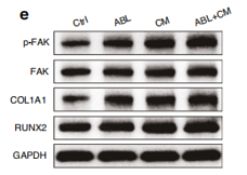
Journal: International Journal Of Oral Science
Application: IHC,WB
Reactivity: Rat
Publish date: 2025 Sept
-
Citation
-
Collagen Glycation-Mediated Mechanical Stress Aggravates Ischemia-Reperfusion injury
Author: Jing Yang, Yixuan Li, Xiaoxiao Fan, Tong Zhang, Junhai Pan, Si Shen, Cheng Zhong, Duguang Li, Yi Zhang, Guoqiao Chen, Wei Ji, Shuqi Wu, Shengxi Jin, Xiaolong Liu, Qiming Xia, Peilin Yu, Wei Yang, Yang Gao, Liangfei Tian, Hui Lin
PMID: 40484296
Journal: Acta Biomaterialia
Application: WB
Reactivity: Human
Publish date: 2025 Jun
-
Citation
-
PYGO1 drives gastric cancer progression via the ITGB1/CD47 axis and is therapeutically targeted by pentagalloylglucose
Author: Jia Yanjuan, Li Yaling, Li Yan, Li Yonghong, Qu Tao, Fu Zhuomin, Ma Yuanyuan, Li Zhenhao, Wang Wanxia, Yu Miao, Jin Xiaojie, Gao Xiaoling, Liu Yongqi
PMID: 40731404
Journal: Journal Of Translational Medicine
Application: WB
Reactivity: Human
Publish date: 2025 Jul
-
Citation
-
Molecular modeling aided design, synthesis and biological evaluation of quinazoline derivatives for the treatment of human cancer
Author: Liu Cai-Shi, Tong Jin-Peng, Fang Ze-Yu, Guo Xiao-Meng, Shi Ting-Ting, Liu Shou-Rong, Sun Juan
PMID: 39832083
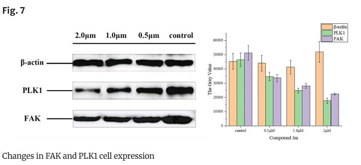
Journal: Molecular Diversity
Application: WB
Reactivity: Human
Publish date: 2025 Jan
-
Citation
-
Dietary biotin facilitates collagen deposition and myofiber growth and development, improving muscle chewiness and hardness in grass carp (Ctenopharyngodon idella)
Author: Yang Yang, Wei-Dan Jiang, Pei Wu, Yang Liu, Yao-Bin Ma, Sheng-Yao Kuang, Shu-Wei Li, Ling Tang, Cheng-Bo Zhong, Lin Feng, Xiao-Qiu Zhou
PMID: NOPMID25100901
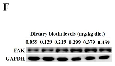
Journal: Food Bioscience
Application: IF,WB
Reactivity: Fish
Publish date: 2025 Aug
-
Citation
-
Integrin β5 interacts with G3BP1 through activating FAK/Src signaling pathway to promote gastric carcinogenesis
Author: Liu Dongliang, Cao Jianjian, Hu Kongwang, Peng Zhiwei
PMID: 40764649
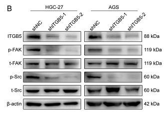
Journal: Scientific Reports
Application: WB
Reactivity: Human
Publish date: 2025 Aug
-
Citation
-
Enhanced migration and adhesion protein expression by polyethylene glycol 4-modified SVVYGLR peptide in an in vitro human gingival fibroblast wound healing model
Author: Peiying Xiong Chengcheng Yu Zhihui Chen Luyuan Chen Yonglong Hong Buling Wu
PMID: 40421822
Journal: Folia Histochemica Et Cytobiologica
Application: IF,WB
Reactivity: Human
Publish date: 2025 Apr
-
Citation
-
Integrin αM promotes macrophage alternative M2 polarization in hyperuricemia-related chronic kidney disease
Author: Jing Liu, Fan Guo, Xiaoting Chen, Ping Fu, Liang Ma
PMID: 38911067
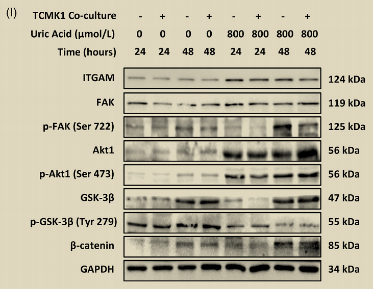
Journal: Medicine And Communication
Application: WB
Reactivity: Mouse
Publish date: 2024 Jun
-
Citation
-
IKIP downregulates THBS1/FAK signaling to suppress migration and invasion by glioblastoma cells
Author: Zhu Zhaoying,et al
PMID: 38948026
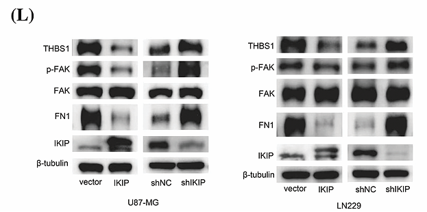
Journal: Oncology Research
Application: WB
Reactivity: Human
Publish date: 2024 Jul
-
Citation
-
Pasteurella multocida activates apoptosis via the FAK-AKT-FOXO1 axis to cause pulmonary integrity loss, bacteremia, and eventually a cytokine storm
Author: Zhao Guangfu,et al
PMID: 38589976
Journal: Veterinary Research
Application: WB
Reactivity: Mouse
Publish date: 2024 Apr
-
Citation
-
PIN1P1 is activated by CREB1 and promotes gastric cancer progression via interacting with YBX1 and upregulating PIN1
Author: Ya-Wen Wang, Wen-Jie Zhu, Ran-Ran Ma, Ya-Ru Tian, Xu Chen, Peng Gao
PMID: 37929660
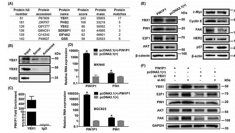
Journal: Journal Of Cellular And Molecular Medicine
Application: WB
Reactivity: Human
Publish date: 2023 Nov
-
Citation
-
Design, synthesis and biological evaluation of novel FAK inhibitors with better selectivity over IR than TAE226
Author:
PMID: 35452915
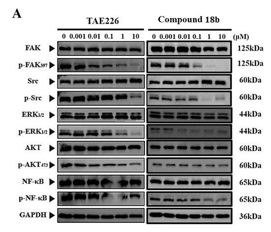
Journal: Bioorganic Chemistry
Application: WB
Reactivity: Human
Publish date: 2022 Jul
-
Citation
-
Ang II-AT2R increases mesenchymal stem cell migration by signaling through the FAK and RhoA/Cdc42 pathways in vitro
Author: Hai-bo Qiu
PMID: 28697804
Journal: Stem Cell Research & Therapy
Application: WB
Reactivity: Human
Publish date: 2017 Jul
-
Citation
Products with the same target and pathway
FAK Rabbit Polyclonal Antibody
Application: WB,IP,ICC,IHC-P
Reactivity: Human,Mouse,Rat
Conjugate: unconjugated
Phospho-FAK (Y397) Recombinant Rabbit Monoclonal Antibody [SC54-07] - BSA and Azide free
Application: WB,IF-Cell,IF-Tissue,IHC-P
Reactivity: Human,Mouse,Rat
Conjugate: unconjugated
FAK Recombinant Rabbit Monoclonal Antibody [SR46-04] - BSA and Azide free
Application: WB,IF-Cell,IF-Tissue,IHC-P,FC,IP
Reactivity: Human,Mouse,Rat
Conjugate: unconjugated
Phospho-FAK (Y397) Recombinant Rabbit Monoclonal Antibody [SC54-07]
Application: WB,IF-Cell,IF-Tissue,IHC-P
Reactivity: Human,Mouse,Rat
Conjugate: unconjugated
Phospho-FAK (S722) Mouse Monoclonal Antibody [4G1]
Application: WB,IP,IF,IHC-P
Reactivity: Human,Mouse,Rat
Conjugate: unconjugated


