Mannose Receptor(CD206) Recombinant Rabbit Monoclonal Antibody [PSH07-76]
Specification
Overview
Product Name
Mannose Receptor(CD206) Recombinant Rabbit Monoclonal Antibody [PSH07-76]
Antibody Type
Recombinant Rabbit monoclonal Antibody
Immunogen
Recombinant protein within mouse Mannose Receptor(CD206) aa 1-1,409.
Species Reactivity
Mouse, Rat
Validated Applications
WB, IHC-P, IF-Tissue, IHC-Fr, IF-Cell, IP, mIHC
Molecular Weight
Predicted band size: 165 kDa
Positive Control
RAW264.7 treated with 20ng/mL mIL-4 and 10ng/mL mIL-13 for 24 hours cell lysate, Mouse lung tissue lysate, Mouse spleen tissue lysate, Rat brain tissue lysate, Rat lung tissue lysate, Rat liver tissue lysate, mouse spleen tissue, mouse liver tissue, rat liver tissue, mouse osteosarcoma tissue.
Conjugation
unconjugated
Clone Number
PSH07-76
Product Features
Form
Liquid
Storage Instructions
Shipped at 4℃. Store at +4℃ short term (1-2 weeks). It is recommended to aliquot into single-use upon delivery. Store at -20℃ long term.
Storage Buffer
PBS (pH7.4), 0.1% BSA, 40% Glycerol. Preservative: 0.05% Sodium Azide.
Isotype
IgG
Purification Method
Protein A affinity purified.
Application Dilution
-
WB
-
1:1,000-1:2,000
-
IHC-P
-
1:1,000
-
IF-Tissue
-
1:500
-
IHC-Fr
-
1:500-1:1,000
-
IF-Cell
-
1:50
-
IP
-
1-2μg/sample
-
mIHC
-
1:2,000
Applications in Publications
| IF | See 7 publications below |
| WB | See 4 publications below |
| IF-cell | See 2 publications below |
| IHC-P | See 1 publications below |
| IHC | See 1 publications below |
| IF-Cell | See 1 publications below |
Species in Publications
Target
Function
The mannose receptor (Cluster of Differentiation 206, CD206) is a C-type lectin primarily present on the surface of macrophages, immature dendritic cells and liver sinusoidal endothelial cells, but is also expressed on the surface of skin cells such as human dermal fibroblasts and keratinocytes. It is the first member of a family of endocytic receptors that includes Endo180 (CD280), M-type PLA2R, and DEC-205 (CD205). The receptor recognises terminal mannose, N-acetylglucosamine and fucose residues on glycans attached to proteins found on the surface of some microorganisms, playing a role in both the innate and adaptive immune systems. Additional functions include clearance of glycoproteins from circulation, including sulphated glycoprotein hormones and glycoproteins released in response to pathological events. The mannose receptor recycles continuously between the plasma membrane and endosomal compartments in a clathrin-dependent manner.
Background References
1. Nielsen MC et al. Macrophage Activation Markers, CD163 and CD206, in Acute-on-Chronic Liver Failure. Cells. 2020 May
2. Nawaz A et al. Depletion of CD206(+) M2-like macrophages induces fibro-adipogenic progenitors activation and muscle regeneration. Nat Commun. 2022 Nov
Subcellular Location
Endosome membrane, Cell membrane.
Synonyms
bA541I19.1 antibody
C type lectin domain family 13 member D antibody
C-type lectin domain family 13 member D antibody
CD 206 antibody
CD206 antibody
CD206 antigen antibody
CLEC13D antibody
CLEC13DL antibody
Macrophage mannose receptor 1 antibody
Macrophage mannose receptor 1 like protein 1 antibody
ExpandbA541I19.1 antibody
C type lectin domain family 13 member D antibody
C-type lectin domain family 13 member D antibody
CD 206 antibody
CD206 antibody
CD206 antigen antibody
CLEC13D antibody
CLEC13DL antibody
Macrophage mannose receptor 1 antibody
Macrophage mannose receptor 1 like protein 1 antibody
Macrophage mannose receptor antibody
Mannose receptor C type 1 antibody
Mannose receptor C type 1 like 1 antibody
MMR antibody
MRC 1 antibody
MRC1 antibody
MRC1_HUMAN antibody
MRC1L1 antibody
OTTHUMP00000045206 antibody
CollapseImages
-

☑ Cell treatment (CT)
Western blot analysis of Mannose Receptor(CD206) on different lysates with Rabbit anti-Mannose Receptor(CD206) antibody (HA722892) at 1/2,000 dilution.
Lane 1: RAW264.7 cell lysate
Lane 2: RAW264.7 treated with 20ng/mL mIL-4 and 10ng/mL mIL-13 for 24 hours cell lysate (Macrophage M2 polarization, positive)
Lane 3: RAW264.7 cell lysate
Lane 4: RAW264.7 treated with 0.1μg/mL LPS for 6 hours cell lysate (Macrophage M1 polarization, negative)
Lysates/proteins at 20 µg/Lane.
Predicted band size: 165 kDa
Observed band size: 200 kDa
Exposure time: 59 seconds; ECL: K1801;
4-20% SDS-PAGE gel.
Proteins were transferred to a PVDF membrane and blocked with 5% NFDM/TBST for 1 hour at room temperature. The primary antibody (HA722892) at 1/2,000 dilution was used in 5% NFDM/TBST at 4℃ overnight. Goat Anti-Rabbit IgG - HRP Secondary Antibody (HA1001) at 1/50,000 dilution was used for 1 hour at room temperature. -

Western blot analysis of Mannose Receptor(CD206) on different lysates with Rabbit anti-Mannose Receptor(CD206) antibody (HA722892) at 1/1,000 dilution.
Lane 1: Mouse lung tissue lysate (20 µg/Lane)
Lane 2: Mouse spleen tissue lysate (20 µg/Lane)
Lane 3: Rat brain tissue lysate (20 µg/Lane)
Lane 4: Rat lung tissue lysate (20 µg/Lane)
Lane 5: Rat liver tissue lysate (20 µg/Lane)
Predicted band size: 165 kDa
Observed band size: 200 kDa
Exposure time: 42 seconds; ECL: K1801;
4-20% SDS-PAGE gel.
Proteins were transferred to a PVDF membrane and blocked with 5% NFDM/TBST for 1 hour at room temperature. The primary antibody (HA722892) at 1/1,000 dilution was used in 5% NFDM/TBST at 4℃ overnight. Goat Anti-Rabbit IgG - HRP Secondary Antibody (HA1001) at 1/50,000 dilution was used for 1 hour at room temperature. -

Application: IHC-Fr
Species: Mouse
Site: Spleen
Sample: Frozen section
Antibody concentration: 1/1,000
Antigen retrieval: Not required -

Application: IHC-Fr
Species: Mouse
Site: Liver
Sample: Frozen section
Antibody concentration: 1/500
Antigen retrieval: Not required -

Immunohistochemical analysis of paraffin-embedded mouse spleen tissue with Rabbit anti-Mannose Receptor(CD206) antibody (HA722892) at 1/1,000 dilution.
The section was pre-treated using heat mediated antigen retrieval with Tris-EDTA buffer (pH 9.0) for 20 minutes. The tissues were blocked in 1% BSA for 20 minutes at room temperature, washed with ddH2O and PBS, and then probed with the primary antibody (HA722892) at 1/1,000 dilution for 1 hour at room temperature. The detection was performed using an HRP conjugated compact polymer system. DAB was used as the chromogen. Tissues were counterstained with hematoxylin and mounted with DPX. -

Immunohistochemical analysis of paraffin-embedded mouse liver tissue with Rabbit anti-Mannose Receptor(CD206) antibody (HA722892) at 1/1,000 dilution.
The section was pre-treated using heat mediated antigen retrieval with Tris-EDTA buffer (pH 9.0) for 20 minutes. The tissues were blocked in 1% BSA for 20 minutes at room temperature, washed with ddH2O and PBS, and then probed with the primary antibody (HA722892) at 1/1,000 dilution for 1 hour at room temperature. The detection was performed using an HRP conjugated compact polymer system. DAB was used as the chromogen. Tissues were counterstained with hematoxylin and mounted with DPX. -

Immunohistochemical analysis of paraffin-embedded rat liver tissue with Rabbit anti-Mannose Receptor(CD206) antibody (HA722892) at 1/1,000 dilution.
The section was pre-treated using heat mediated antigen retrieval with Tris-EDTA buffer (pH 9.0) for 20 minutes. The tissues were blocked in 1% BSA for 20 minutes at room temperature, washed with ddH2O and PBS, and then probed with the primary antibody (HA722892) at 1/1,000 dilution for 1 hour at room temperature. The detection was performed using an HRP conjugated compact polymer system. DAB was used as the chromogen. Tissues were counterstained with hematoxylin and mounted with DPX. -

☑ Cell treatment (CT)
Immunocytochemistry analysis of RAW264.7 cells treated with 20ng/mL mIL-4 and 10ng/mL mIL-13 for 24 hours (Macrophage M2 polarization, positive) labeling Mannose Receptor(CD206) with Rabbit anti-Mannose Receptor(CD206) antibody (HA722892) at 1/50 dilution.
Cells were fixed in 4% paraformaldehyde for 15 minutes at room temperature, permeabilized with 0.1% Triton X-100 in PBS for 15 minutes at room temperature, then blocked with 1% BSA in 10% negative goat serum for 1 hour at room temperature. Cells were then incubated with Rabbit anti-Mannose Receptor(CD206) antibody (HA722892) at 1/50 dilution in 1% BSA in PBST overnight at 4 ℃. Goat Anti-Rabbit IgG H&L (iFluor™ 488, HA1121) was used as the secondary antibody at 1/1,000 dilution. PBS instead of the primary antibody was used as the secondary antibody only control. Nuclear DNA was labelled in blue with DAPI.
Beta tubulin (M1305-2, red) was stained at 1/100 dilution overnight at +4℃. Goat Anti-Mouse IgG H&L (iFluor™ 594, HA1126) was used as the secondary antibody at 1/1,000 dilution. -

☑ Cell treatment (CT)
Immunocytochemistry analysis of RAW264.7 cells treated with 0.1μg/mL LPS for 6 hours (Macrophage M1 polarization, negative) labeling Mannose Receptor(CD206) with Rabbit anti-Mannose Receptor(CD206) antibody (HA722892) at 1/50 dilution.
Cells were fixed in 4% paraformaldehyde for 15 minutes at room temperature, permeabilized with 0.1% Triton X-100 in PBS for 15 minutes at room temperature, then blocked with 1% BSA in 10% negative goat serum for 1 hour at room temperature. Cells were then incubated with Rabbit anti-Mannose Receptor(CD206) antibody (HA722892) at 1/50 dilution in 1% BSA in PBST overnight at 4 ℃. Goat Anti-Rabbit IgG H&L (iFluor™ 488, HA1121) was used as the secondary antibody at 1/1,000 dilution. PBS instead of the primary antibody was used as the secondary antibody only control. Nuclear DNA was labelled in blue with DAPI.
Beta tubulin (M1305-2, red) was stained at 1/100 dilution overnight at +4℃. Goat Anti-Mouse IgG H&L (iFluor™ 594, HA1126) was used as the secondary antibody at 1/1,000 dilution. -

Application: IF-tissue
Species: Mouse
Site: Spleen
Sample: Paraffin-embedded section
Antibody concentration: 1/500 -

Application: IF-tissue
Species: Mouse
Site: Liver
Sample: Paraffin-embedded section
Antibody concentration: 1/500 -

Mannose Receptor(CD206) was immunoprecipitated from 0.2 mg rat lung tissue lysate with HA722892 at 2 µg/10 µl beads. Western blot was performed from the immunoprecipitate using HA722892 at 1/1,000 dilution. Anti-Rabbit IgG for IP Nano-secondary antibody (NBI01H) at 1/5,000 dilution was used for 1 hour at room temperature.
Lane 1: rat lung tissue lysate (input)
Lane 2: HA722892 IP in rat lung tissue lysate
Lane 3: Rabbit IgG instead of HA722892 in rat lung tissue lysate
Blocking/Dilution buffer: 5% NFDM/TBST
Exposure time: 1 minute; ECL: K1801 -

mIHC analysis of mouse liver tissue (Formalin/PFA-fixed paraffin-embedded sections) with Rabbit anti-Mannose Receptor(CD206) antibody (HA722892) at 1/2,000 dilution. The immunostaining was performed with the IRISKitCmTSA Kit (900808). Heat mediated antigen retrieval with Tris-EDTA buffer (pH 9.0) for 30 mins at 95℃. DAPI (blue) was used as a nuclear counter stain. Image acquisition was performed with Olympus VS200 Slide Scanner.
-

mIHC analysis of mouse osteosarcoma tissue (Formalin/PFA-fixed paraffin-embedded sections) with Rabbit anti-Mannose Receptor(CD206) antibody (HA722892) at 1/2,000 dilution. The immunostaining was performed with the IRISKitCmTSA Kit (900808). Heat mediated antigen retrieval with Tris-EDTA buffer (pH 9.0) for 30 mins at 95℃. DAPI (blue) was used as a nuclear counter stain. Image acquisition was performed with Olympus VS200 Slide Scanner.
-

mIHC analysis of mouse spleen tissue (Formalin/PFA-fixed paraffin-embedded sections) with Rabbit anti-Mannose Receptor(CD206) antibody (HA722892) at 1/2,000 dilution. The immunostaining was performed with the IRISKitCmTSA Kit (900808). Heat mediated antigen retrieval with Tris-EDTA buffer (pH 9.0) for 30 mins at 95℃. DAPI (blue) was used as a nuclear counter stain. Image acquisition was performed with Olympus VS200 Slide Scanner.
Please note: All products are "FOR RESEARCH USE ONLY AND ARE NOT INTENDED FOR DIAGNOSTIC OR THERAPEUTIC USE"
Citation
-
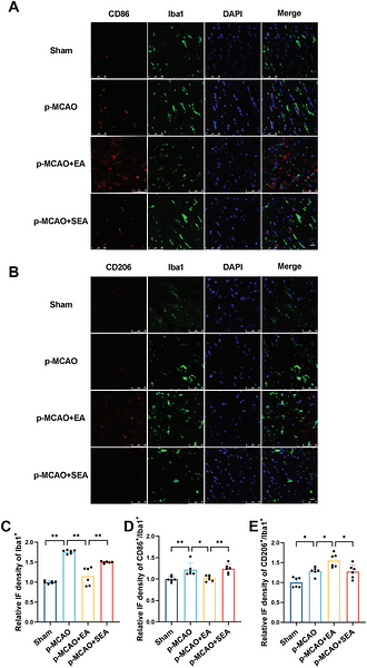
Electroacupuncture promotes myelin repair in the corpus callosum region of cerebral ischemic rats by modulating microglia activation and polarization
Author: Feng Wu
PMID: 40923850

Journal: Neurological Research
Application: WB
Reactivity: Rat
Publish date: 2025 Sept
-
Citation
-
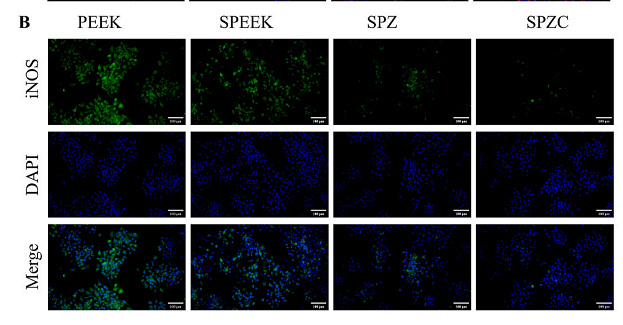
Fennel essential oil and its nanoemulsion modulate macrophage-mediated inflammatory responses and promote pressure ulcer healing
Author: Zhiling Luo, Liying Zeng, Zhilin Zhao, Yulian Zheng, Dong Wan
PMID: 41101230
Journal: International Immunopharmacology
Application: IF-cell
Reactivity: Mouse,Rat
Publish date: 2025 Oct
-
Citation
-
Brain abundant membrane attached signal protein 1 regulates macrophage polarization through EGFR/PI3K/AKT signaling in renal cell carcinoma
Author: Taian Jin, Luping Wang, Ruikai Zhang, Mengting Wu, Xinyang Fang, Binqi Wang, Wenfang He, Weiwei Qian, Juan Jin, Qiang He
PMID: 41093214
Journal: International Journal Of Biological Macromolecules
Application: WB
Reactivity: Mouse
Publish date: 2025 Oct
-
Citation
-
Silicified strontium-curcumin chelated nanospheres mitigate inflammatory vicious cycles to accelerate diabetic wound healing
Author: Zhiwei Zhao, Youjun Ding, Guangjin Gao, Yepeng Zhang, Zhaowenbin Zhang, Min Zhou, Ye Yuan
PMID: 41142414
Journal: Materials Today Bio
Application: WB
Reactivity: Mouse
Publish date: 2025 Oct
-
Citation
-
Hepatic stellate cells shape the ECM-disorganized and immunosuppressive microenvironment via CCL11/CCR3 axis in lenvatinib-treated hepatocellular carcinoma
Author: Na Qiang, Cheng Lan, An Lin, Hiroaki Kanzaki, Masanori Inoue, Naoya Kanogawa, Takayuki Kondo, Sadahisa Ogasawara, Masato Nakamura, Shingo Nakamoto, Dawei Cui, Hang Lv, Qiuran Xu, Guiping Chen, Junjie Ao
PMID: 10.1016/j.cellsig.2025.112263
Journal: Cellular Signalling
Application: IHC
Reactivity: Mouse
Publish date: 2025 Nov
-
Citation
-
Brain-derived extracellular vesicles circUsp32 polarized macrophages causing acute kidney injury after traumatic brain injury
Author: Jiayuanyuan Fu, Jingheng Wu, Mengran Du, Xu Wang, Qi Shi, Yehong Fang, Yu Lan, Qiaoli Wu, Guobin Zhang, Lixia Xu, Hua Yan
PMID: 10.1126/sciadv.adz1243
Journal: Science Advances
Application: IF
Reactivity: Mouse
Publish date: 2025 Nov
-
Citation
-
An Injectable Hydrogel Enabling Photo-Temporal Modulation of Asymmetric Adhesion for Tissue Fixation and Prevention of Postoperative Peritoneal Adhesion
Author: Zhongming Zhao, Xiang Ji, Xiuqiang Li, Jiazhuo Gong, Yi Gao, Zhenhao Li, Mengmeng Yao, Chaojie Yu, Fanglian Yao, Hong Sun, Junjie Li, Hong Zhang
PMID: 10.1002/adfm.202516756
Journal: Advanced Functional Materials
Application: IF-cell
Reactivity: Mouse
Publish date: 2025 Nov
-
Citation
-
Curcumin encapsulated zeolitic imidazolate frameworks modified polyetheretherketone promotes osteogenic capacity through immunomodulation
Author: Sihan Ma, Heyi Gong, Duo Sun, Xin Fang, Xiao Han, Yue Liu, Yadi Su, Junqi Wang, Yu Wang, Jinghui Zhao
PMID: 19NOPMID25051201

Journal: Composites Science and Technology
Application: IF
Reactivity: Mouse
Publish date: 2025 Mar
-
Citation
-
High mobility group box 1, a novel serotonin receptor-7 negative modulator, contributes to M2 microglial ferroptosis and neuroinflammation in post-stroke depression
Author: Ou Du, Chao Wu, Yu-Xin Yang, Han-Yinan Yang, Yi-Jin Wu, Meng-Yang Li, Si-cen Liu, Zhen-Hua Shao, Jun-Rong Du
PMID: 40527447
Journal: Free Radical Biology And Medicine
Application: IF
Reactivity: Mouse
Publish date: 2025 Jun
-
Citation
-
A Zinc-Citrate Metal–Organic Framework-Based Adaptable Hydrogen Sulfide Delivery System for Regulating Neuroregeneration Microenvironment in Spinal Cord Injury
Author: Yawei Yao, Xianzhen Dong, Zixuan Pang, Jingyuan Shao, Zhichao He, Kun Liu, Peihong Hou, Fanqi Hu, Weibo Liu, Yuanfang Huo, Hua Wang, Honglian Dai, Xuesong Zhang
PMID: 40524455
Journal: ACS Applied Nano Materials
Application: WB
Reactivity: Mouse,Rat
Publish date: 2025 Jun
-
Citation
-
A wearable spatiotemporal controllable ultrasonic device with amyloid-β disaggregation for continuous Alzheimer’s disease therapy
Author:
PMID: 40737409
Journal: Science Advances
Application: IF
Reactivity: Mouse
Publish date: 2025 Jul
-
Citation
-
Incorporating Stem Cell-Derived Small Extracellular Vesicles into a Multifunctional OHAMA-EPL Hydrogel Spray for Rapid Healing of Infected Skin Wounds
Author: Mingzhu He, Yongchun Wei, Xujing Chen, Jingqi Li, Jingjing Kang, Bowei Li, Dingbin Liu, Hong Cai
PMID: 40697065
Journal: Advanced Healthcare Materials
Application: IF
Reactivity: Mouse
Publish date: 2025 Jul
-
Citation
-
SZC-6 Promotes Diabetic Wound Healing in Mice by Modulating the M1/M2 Macrophage Ratio and Inhibiting the MyD88/NF-χB Pathway
Author: Ang Xuan, Meng Liu, Lingli Zhang, Guoqing Lu, Hao Liu, Lishan Zheng, Juan Shen, Yong Zou, Shengyao Zhi
PMID: 25NOPMID25082602
Journal: Pharmaceuticals
Application: IF
Reactivity: Mouse
Publish date: 2025 Jul
-
Citation
-
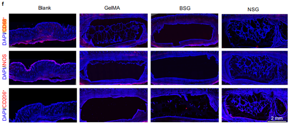
Peptide-based rigid nanorod-reinforced gelatin methacryloyl hydrogels for osteochondral regeneration and additive manufacturing
Author: Zhu Junjin, Wei Yueting, Yang Guangmei, Zhang Jiangnan, Liu Jiayi, Zhang Xin, Zhu Zhou, Chen Junyu, Pei Xibo, Wu Dongdong, Wang Jian
PMID: 40750776

Journal: Nature Communications
Application: IF
Reactivity: Rat
Publish date: 2025 Aug
-
Citation
-
Lumbrokinase-loaded GelMA hydrogels with inflammatory regulatory capacity promote vascularized bone regeneration in critical-sized cranial defects
Author:
PMID: 10.1039/D5RA04178C
Journal: RSC Advances
Application: IF-Cell
Reactivity: Mouse
Publish date: 2025 Aug
-
Citation
-
2A-biohydrogels accelerate diabetic wound healing by promoting M2 macrophage polarization and functionalized mitochondrial transfer to endothelial cells
Author: Hao Wang, Min Zhou, Yifeng Ruan, Yifei Shen, Ziqi Qin, Xingbo Wu, Huiling Ling, Wushuang Ye, Yongfu Wang, Xueqi Gan
PMID: 12NOPMID25052801
Journal: Chemical Engineering Journal
Application: IHC-P
Reactivity: Rat
Publish date: 2025 Apr
-
Citation

