Autophagy Marker Antibody Sampler Kit


Catalog# HAK21168
Autophagy Marker Antibody Sampler Kit
-
WB
-
Human
-
Mouse
Overview
Kit Components
| Product Includes | Specification | Application | Reactivity | Mw |
|---|---|---|---|---|
| ATG5[ET1611-38] | 20µl | WB,IF-Cell,IF-Tissue,IHC-P,IP,FC | Human,Mouse,Rat,Monkey | Predicted band size: 32 kDa |
| ATG16L1[ET7106-65] | 20µl | WB,IHC-P | Human,Mouse,Rat | Predicted band size: 68 kDa |
| ATG4B[HA721829] | 20µl | WB | Human,Mouse,Rat,Monkey | Predicted band size: 44 kDa |
| LC3B[ET1701-65] | 20µl | WB,IF-Cell,IHC-P,IF-Tissue,IP,mIHC | Human,Mouse,Rat | Predicted band size: 14/16 kDa |
| ATG9A[ET1610-71] | 20µl | WB,IHC-P,IP | Human,Mouse,Rat | Predicted band size: 94 kDa |
| Beclin 1[HA721216] | 20µl | WB,IHC-P,IF-Cell,FC | Human,Mouse,Rat | Predicted band size: 52 kDa |
| Goat Anti-Rabbit IgG (H+L)[HA1001] | 100µl | WB,ELISA,IHC-P | Rabbit |
Product Description
The Autophagy Marker Antibody Sampler Kit provides an economical means of detecting proteins related to autophagy. The kit contains enough antibody to perform two western blot experiments per primary antibody.
Product Features
Storage Buffer
1*TBS (pH7.4), 0.05% BSA, 40% Glycerol. Preservative: 0.05% Sodium Azide.
Storage Instructions
Store at +4℃ after thawing. Aliquot store at -20℃. Avoid repeated freeze / thaw cycles.
Background
Autophagy is a highly dynamic process consisting of the following three steps: (1) autophagosome formation, (2) autophagosome-lysosome fusion, and (3) degradation.<br> <br> The strategic combination of antibodies against ATG5, ATG16L1, ATG4B, LC3B, ATG9A, and Beclin 1 constitutes a powerful and complete toolkit for autophagy research. This panel allows researchers to precisely dissect the dynamic process of autophagy at its most critical stages: from Initiation (Beclin 1) and Membrane Sourcing (ATG9A), through Phagophore Elongation (ATG5, ATG16L1), to final Vesicle Maturation & Turnover (LC3B, ATG4B).
Data Links
Background References
1. Liu S, Yao S, Yang H, Liu S, Wang Y. Autophagy: Regulator of cell death. Cell Death Dis. 2023 Oct 4;14(10):648.
2. Glick D, Barth S, Macleod KF. Autophagy: cellular and molecular mechanisms. J Pathol. 2010 May;221(1):3-12.
3. Mizushima N, Komatsu M. Autophagy: renovation of cells and tissues. Cell. 2011 Nov 11;147(4):728-41.
Images
-
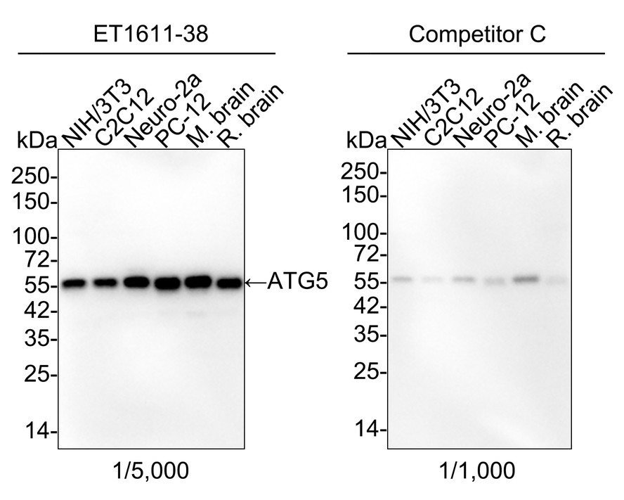
Western blot analysis of ATG5 on different lysates with Rabbit anti-ATG5 antibody (ET1611-38) at 1/5,000 dilution and competitor's antibody at 1/1,000 dilution.
Lane 1: NIH/3T3 cell lysate (15 µg/Lane)
Lane 2: C2C12 cell lysate (15 µg/Lane)
Lane 3: Neuro-2a cell lysate (15 µg/Lane)
Lane 4: PC-12 cell lysate (15 µg/Lane)
Lane 5: Mouse brain tissue lysate (15 µg/Lane)
Lane 6: Rat brain tissue lysate (15 µg/Lane)
Predicted band size: 32 kDa
Observed band size: 55 kDa
Exposure time: 35 seconds; ECL: K1802;
4-20% SDS-PAGE gel.
Proteins were transferred to a PVDF membrane and blocked with 5% NFDM/TBST for 1 hour at room temperature. The primary antibody (ET1611-38) at 1/5,000 dilution and competitor's antibody at 1/1,000 dilution were used in 5% NFDM/TBST at 4℃ overnight. Goat Anti-Rabbit IgG - HRP Secondary Antibody (HA1001) at 1/50,000 dilution was used for 1 hour at room temperature. -
Immunocytochemistry analysis of NIH/3T3 cells labeling ATG5 with Rabbit anti-ATG5 antibody (ET1611-38) at 1/250 dilution.
Cells were fixed in 4% paraformaldehyde for 20 minutes at room temperature, permeabilized with 0.1% Triton X-100 in PBS for 5 minutes at room temperature, then blocked with 1% BSA in 10% negative goat serum for 1 hour at room temperature. Cells were then incubated with Rabbit anti-ATG5 antibody (ET1611-38) at 1/250 dilution in 1% BSA in PBST overnight at 4 ℃. Goat Anti-Rabbit IgG H&L (iFluor™ 488, HA1121) was used as the secondary antibody at 1/1,000 dilution. PBS instead of the primary antibody was used as the secondary antibody only control. Nuclear DNA was labelled in blue with DAPI. Beta tubulin (M1305-2, red) was stained at 1/100 dilution overnight at +4℃. Goat Anti-Mouse IgG H&L (iFluor™ 594, HA1126) was used as the secondary antibody at 1/1,000 dilution. -
Western blot analysis of ATG4B on different lysates with Rabbit anti-ATG4B antibody (HA721334) at 1/5,000 dilution.
Lane 1: 293T cell lysate
Lane 2: MCF7 cell lysate
Lane 3: COS-1 cell lysate
Lane 4: NIH/3T3 cell lysate
Lane 5: PC-12 cell lysate
Lane 6: Mouse brain tissue lysate
Lane 7: Rat brain tissue lysate
Lysates/proteins at 20 µg/Lane.
Predicted band size: 44 kDa
Observed band size: 47 kDa
Exposure time: 20 seconds; ECL: K1801;
4-20% SDS-PAGE gel.
Proteins were transferred to a PVDF membrane and blocked with 5% NFDM/TBST for 1 hour at room temperature. The primary antibody (HA721334) at 1/5,000 dilution was used in primary antibody dilution (K1803) at 4℃ overnight. Goat Anti-Rabbit IgG - HRP Secondary Antibody (HA1001) at 1/50,000 dilution was used for 1 hour at room temperature. -
ATG5 was immunoprecipitated in 0.2mg HeLa cell lysate with ET1611-38 at 2 µg/10 µl beads. Western blot was performed from the immunoprecipitate using ET1611-38 at 1/2,000 dilution. Anti-Rabbit IgG for IP Nano-secondary antibody (NBI01H) at 1/5,000 dilution was used for 1 hour at room temperature.
Lane 1: HeLa cell lysate (input)
Lane 2: Rabbit IgG instead of ET1611-38 in HeLa cell lysate
Lane 3: ET1611-38 IP in HeLa cell lysate
Blocking/Dilution buffer: 5% NFDM/TBST
Exposure time: 1 minute 21 seconds; ECL: K1802 -
Western blot analysis of ATG4B on different lysates with Rabbit anti-ATG4B antibody (HA721829) at 1/2,000 dilution and competitor's antibody at 1/2,000 dilution.
Lane 1: HeLa cell lysate
Lane 2: Jurkat cell lysate
Lane 3: 293T cell lysate
Lane 4: MCF7 cell lysate
Lane 5: K-562 cell lysate
Lane 6: COS-1 cell lysate
Lane 7: NIH/3T3 cell lysate
Lane 8: C2C12 cell lysate
Lane 9: PC-12 cell lysate
Lane 10: Mouse brain tissue lysate
Lane 11: Rat brain tissue lysate
Lane 12: Mouse kidney tissue lysate
Lane 13: Mouse colon tissue lysate
Lysates/proteins at 30 µg/Lane.
Predicted band size: 44 kDa
Observed band size: 47 kDa
Exposure time: 42 seconds; ECL: K1802;
4-20% SDS-PAGE gel.
Proteins were transferred to a PVDF membrane and blocked with 5% NFDM/TBST for 1 hour at room temperature. The primary antibody (HA721829) at 1/2,000 dilution and competitor's antibody at 1/2,000 dilution were used in 5% NFDM/TBST at 4℃ overnight. Goat Anti-Rabbit IgG - HRP Secondary Antibody (HA1001) at 1/50,000 dilution was used for 1 hour at room temperature. -
Immunocytochemistry analysis of C2C12 cells treated with or without 50μM Chloroquine for 24 hours labeling LC3B with Rabbit anti-LC3B antibody (ET1701-65) at 1/100 dilution.
Cells were fixed in 4% paraformaldehyde for 20 minutes at room temperature, permeabilized with 0.1% Triton X-100 in PBS for 5 minutes at room temperature, then blocked with 1% BSA in 10% negative goat serum for 1 hour at room temperature. Cells were then incubated with Rabbit anti-LC3B antibody (ET1701-65) at 1/100 dilution in 1% BSA in PBST overnight at 4 ℃. Goat Anti-Rabbit IgG H&L (iFluor™ 488, HA1121) was used as the secondary antibody at 1/1,000 dilution. PBS instead of the primary antibody was used as the secondary antibody only control. Nuclear DNA was labelled in blue with DAPI. Beta tubulin (M1305-2, red) was stained at 1/100 dilution overnight at +4℃. Goat Anti-Mouse IgG H&L (iFluor™ 594, HA1126) was used as the secondary antibody at 1/1,000 dilution. -
Immunocytochemistry analysis of HeLa cells treated with or without 50μM Chloroquine for 24 hours labeling LC3B with Rabbit anti-LC3B antibody (ET1701-65) at 1/200 dilution.
Cells were fixed in 4% paraformaldehyde for 10 minutes at 37 ℃, permeabilized with 0.05% Triton X-100 in PBS for 20 minutes, and then blocked with 2% negative goat serum for 30 minutes at room temperature. Cells were then incubated with Rabbit anti-LC3B antibody (ET1701-65) at 1/200 dilution in 2% negative goat serum overnight at 4 ℃. Goat Anti-Rabbit IgG H&L (iFluor™ 488, HA1121) was used as the secondary antibody at 1/1,000 dilution. Nuclear DNA was labelled in blue with DAPI.
Beta tubulin (M1305-2, red) was stained at 1/100 dilution overnight at +4℃. Goat Anti-Mouse IgG H&L (iFluor™ 594, HA1126) was used as the secondary antibody at 1/1,000 dilution. -
Western blot analysis of LC3B on different lysates with Rabbit anti-LC3B antibody (ET1701-65) at 1/2,000 dilution.
Lane 1: HCT 116 cell lysate
Lane 2: HCT 116 treated with 50μM Chloroquine for 18 hours cell lysate
Lane 3: U-87 MG cell lysate
Lane 4: C6 cell lysate
Lane 5: Mouse brain tissue lysate
Lane 6: Rat brain tissue lysate
Lysates/proteins at 20 µg/Lane.
Predicted band size: 14/16 kDa
Observed band size: 14/16 kDa
Exposure time: 26 seconds; ECL: K1801;
4-20% SDS-PAGE gel.
Proteins were transferred to a PVDF membrane and blocked with 5% NFDM/TBST for 1 hour at room temperature. The primary antibody (ET1701-65) at 1/2,000 dilution was used in 5% NFDM/TBST at 4℃ overnight. Goat Anti-Rabbit IgG - HRP Secondary Antibody (HA1001) at 1/50,000 dilution was used for 1 hour at room temperature. -
Western blot analysis of Beclin 1 on different lysates with Rabbit anti-Beclin 1 antibody (HA721216) at 1/5,000 dilution.
Lane 1: HeLa cell lysate (20 µg/Lane)
Lane 2: HepG2 cell lysate (20 µg/Lane)
Lane 3: NIH/3T3 cell lysate (20 µg/Lane)
Lane 4: C6 cell lysate (20 µg/Lane)
Lane 5: Mouse cerebellum tissue lysate (40 µg/Lane)
Lane 6: Rat cerebellum tissue lysate (40 µg/Lane)
Predicted band size: 52 kDa
Observed band size: 57 kDa
Exposure time: 10 seconds; ECL: K1801;
4-20% SDS-PAGE gel.
Proteins were transferred to a PVDF membrane and blocked with 5% NFDM/TBST for 1 hour at room temperature. The primary antibody (HA721216) at 1/5,000 dilution was used in primary antibody dilution (K1803) at 4℃ overnight. Goat Anti-Rabbit IgG - HRP Secondary Antibody (HA1001) at 1/50,000 dilution was used for 1 hour at room temperature. -
Immunocytochemistry analysis of HeLa cells labeling Beclin 1 with Rabbit anti-Beclin 1 antibody (HA721216) at 1/100 dilution.
Cells were fixed in 4% paraformaldehyde for 15 minutes at room temperature, permeabilized with 0.1% Triton X-100 in PBS for 15 minutes at room temperature, then blocked with 1% BSA in 10% negative goat serum for 1 hour at room temperature. Cells were then incubated with Rabbit anti-Beclin 1 antibody (HA721216) at 1/100 dilution in 1% BSA in PBST overnight at 4 ℃. Goat Anti-Rabbit IgG H&L (iFluor™ 488, HA1121) was used as the secondary antibody at 1/1,000 dilution. PBS instead of the primary antibody was used as the secondary antibody only control. Nuclear DNA was labelled in blue with DAPI.
Beta tubulin (HA601187, red) was stained at 1/100 dilution overnight at +4℃. Goat Anti-Mouse IgG H&L (iFluor™ 594, HA1126) was used as the secondary antibody at 1/1,000 dilution. -
Flow cytometric analysis of PC-12 cells labeling ATG5.
Cells were fixed and permeabilized. Then stained with the primary antibody (ET1611-38, 1/1,000) (red) compared with Rabbit IgG Isotype Control (green). After incubation of the primary antibody at +4℃ for an hour, the cells were stained with a iFluor™ 488 conjugate-Goat anti-Rabbit IgG Secondary antibody (HA1121) at 1/1,000 dilution for 30 minutes at +4℃. Unlabelled sample was used as a control (cells without incubation with primary antibody; black).
Please note: All products are "FOR RESEARCH USE ONLY AND ARE NOT INTENDED FOR DIAGNOSTIC OR THERAPEUTIC USE"
Related Products

Autophagy Vesicle Elongation (Atg12 Conjugation) Antibody Sampler Kit
Application: WB
Reactivity: Human
Conjugate:
ATG5 Recombinant Rabbit Monoclonal Antibody [SN73-07]
Application: WB,IF-Cell,IF-Tissue,IHC-P,IP,FC
Reactivity: Human,Mouse,Rat,Monkey
Conjugate: unconjugated
